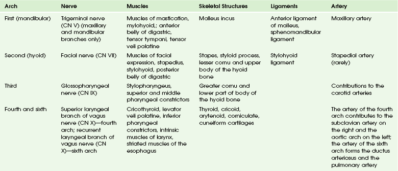
Embryology, Anatomy, Normal Findings, and Imaging Techniques

Chapter 13


Embryology, Anatomy, Normal Findings, and Imaging Techniques


Embryology of the Neck


Accurate diagnosis and successful treatment of congenital anomalies and masses of the neck are dependent on an understanding of the complex embryologic development of this region and the anomalies that result from abnormal development.


This chapter will focus on the embryology of the neck and the oral cavity. The embryology of the orbit, face and sinuses, temporal bone, and ear are addressed in Chapters 4, 8, 9, and 18.


Many of the structures of the head and neck form from an interaction between somitomeres, somites, the mesenchyme, and the branchial apparatus.


Development of the Mesoderm, Somitomeres, and Somites
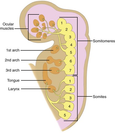

After neurulation occurs, the mesoderm subdivides into the lateral, intermediate, and paraxial mesoderm. The lateral mesoderm forms most of the throat and larynx. The intermediate mesoderm does not form any part of the head and neck. The paraxial mesoderm forms the seven somitomeres and 42 to 44 paired somites. The five most rostral somites are involved in the formation of head and neck musculature (Fig. 13-1). The somitomeres and somites form before the development of the branchial apparatus.


The branchial apparatus, that is, the branchial arches, clefts, pouches, and membrane, begin to form late in the third week of gestation. The buccopharyngeal membrane breaks down and the mesodermal branchial bars begin to form six pairs of branchial arches. The fifth arch is rudimentary and disappears.


The fourth somitomere invades the first branchial arch and generates the formation of the muscles of mastication, that is, the masseter, pterygoid, and temporalis muscles. These muscles are innervated by the trigeminal nerve (CN V).


The seventh somitomere interacts with the third brachial arch to form the stylopharyngeus muscle, which is innervated by the glossopharyngeal nerve (CN IX).


The first four occipital somites invade the fourth and sixth brachial arches and thus stimulate the formation of the extrinsic and intrinsic laryngeal muscles innervated by the vagus nerve (CN X) and the cranial segment of the spinal accessory nerve (CN XI).


The third through seventh somites form the sternocleidomastoid muscle and trapezius and are innervated by the spinal accessory nerve (CN XI).


The intrinsic and extrinsic tongue muscles are likely derived from the second through fourth occipital somites and are innervated by the hypoglossal nerve (CN XII).


The contribution of the somitomeres and somites to the formation of muscles and their distinct innervation is unchanged throughout growth and development. Thus although many muscles migrate in location, their nerve supply is maintained and hence their branchial arch origin can always be identified (see Fig. 13-1).


Development of the Branchial Apparatus


Formation of the branchial apparatus occurs between the fourth through seventh weeks of development. The pharynx constitutes much of the foregut during the first few weeks of development. Formation of the five branchial arches (I, II, III, IV, and VI) results from the breakdown of the buccopharyngeal membrane and segmentation of the mesoderm. Migration of neural crest cells to this location stimulates growth and development. Each arch has its own outer epithelial lining of ectoderm separated by five clefts and an inner epithelial lining of endoderm with five corresponding pouches and a central cartilaginous core, which is a mesenchymal derivative that participates in the formation of the characteristic skeletal, muscular, ligamentous, vascular, and neural components of each arch.


Shortly after formation of the branchial arches, the first and second arches undergo mesodermal proliferation, thus creating the epicardial ridge, which contains the mesodermal precursor of the sternocleidomastoid, the trapezius, and the infrahyoid and lingual muscles. The nerves of the epicardial ridge are the hypoglossal (CN XII) and spinal accessory (CN XI) nerves. The proliferation of mesenchyme overgrows branchial arches II, III, and IV and narrows branchial clefts II, III, and IV. Subsequently, an ectodermal pit is formed—the cervical sinus of His—which obliterates with further development; failure of obliteration results in formation of branchial sinus, clefts, or cysts of types II, III, or IV.


Branchial Apparatus and its Contribution to the Structures of the Neck
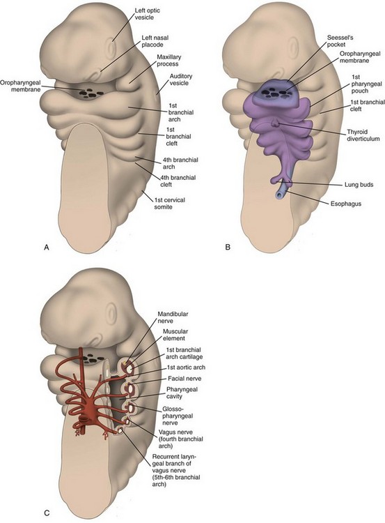


Branchial Arches: The first branchial arch (Fig. 13-2) is composed of a dorsal segment known as the maxillary process and a ventral segment known as Meckel cartilage or the mandibular process; both involute. The ossification around Meckel cartilage is the precursor of the mandible and the sphenomandibular cartilage in the neck. The muscle derivatives of the first arch are the muscles of mastication (the masseter, pterygoid, and temporalis muscles), the tensor tympani and tensor veli palatine muscles, the anterior belly of the digastric muscle, and the mylohyoid muscle. The trigeminal nerve (CN V) provides motor and sensory innervation to the first branchial arch.


The second branchial arch is also known as Reichert cartilage. It gives rise to the upper body and lesser cornu of the hyoid bone, the styloid process, and stylohyoid ligament. The muscle derivatives include the platysma, the posterior belly of the digastric, and the stylohyoid. The nerve of the second brachial arch is the facial nerve (CN VII), which is primarily motor. The main sensory component is the chorda tympani branch that is carried with a branch of the trigeminal nerve (CN V3) to supply taste to the anterior two thirds of the tongue. The artery of the second brachial arch is the stapedial artery, which normally regresses aside from some contributions to the internal and external carotid arteries.


The third branchial arch cartilage derivatives include the greater cornu and inferior body of the hyoid. The muscle derivatives include the stylopharyngeus and superior and middle pharyngeal constrictors. The nerve of the third brachial arch is the glossopharyngeal nerve (CN IX). The neural crest cells of the third branchial arch also form the carotid bodies. The artery of the third branchial arch contributes to the common carotid artery and the internal and external carotid arteries.


The fourth and sixth branchial arch cartilage derivatives fuse to form the larynx and the laryngeal cartilages (the thyroid, cricoid, arytenoid, corniculate, and cuneiform). Muscle derivatives include the cricothyroid muscle, the levator veli palatini, and the inferior pharyngeal constrictors. The muscle derivatives of the sixth arch are the remaining intrinsic muscles of the larynx. The nerve of the fourth arch is the superior laryngeal nerve, and the nerve of the sixth arch is the recurrent laryngeal nerve. Both are branches of the vagus nerve (CN X). The artery of the fourth branchial arch contributes to the aortic arch on the left and the subclavian artery on the right. The artery of the sixth branchial arch becomes the ductus arteriosus and the pulmonary artery. Between the branchial arches lie the paired branchial pouches and clefts.


Branchial Pouches: The first branchial pouch does not contribute to the structures of the neck. The second branchial pouch gives rise to the palatine tonsils and tonsillar fossa. The third branchial pouch gives rise to the inferior parathyroids and thymus. The early embryologic connections to the pharynx normally are obliterated. The fourth branchial pouch gives rise to the superior parathyroid glands and the ultimobranchial body, which contains the parafollicular cells (C cells) of the thyroid gland. The fifth branchial pouch degenerates. The branchial clefts do not contribute to any neck structures and are obliterated as development occurs (Tables 13-1 and 13-2).


Table 13-2


Derivatives of the Branchial Pouches


Only gold members can continue reading. Log In or Register to continue

Stay updated, free articles. Join our Telegram channel

Dec 20, 2015 | Posted by in PEDIATRIC IMAGING | Comments Off on Embryology, Anatomy, Normal Findings, and Imaging Techniques

Full access? Get Clinical Tree

Get Clinical Tree app for offline access
Pouch Derivatives
First Eustachian tube, middle ear, portions of mastoid bone
Second Palatine tonsils, tonsillar fossa
Third Inferior parathyroids, thymus
Fourth and sixth Superior parathyroids, parafollicular cells of thyroid