Jean Noel Buy1 and Michel Ghossain2
(1)
Service Radiologie, Hopital Hotel-Dieu, Paris, France
(2)
Department of Radiology, Hotel Dieu de France, Beirut, Lebanon
Abstract
The urogenital system develops from the intermediate mesenchyme derived from the dorsal body wall of the embryo (Fig. 1.1).
1.1 Embryology of the Urogenital System
The urogenital system develops from the intermediate mesenchyme derived from the dorsal body wall of the embryo (Fig. 1.1).
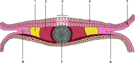

Fig. 1.1
1 Intermediate mesenchyme (mesoderm), 2 paraxial mesoderm, 3 neural groove, 4 embryonic ectoderm, 5 amnion, 6 coelomic spaces, 7 notochord, 8 lateral mesoderm [Adapted from Moore [1]]
At the beginning of the third week, an opacity formed by a thickened linear band of epiblast—the primitive streak—appears caudally in the median plane of the dorsal aspect of the embryonic disc.
Shortly after the primitive streak appears, cells leave its deep surface and form mesenchyme, a tissue consisting of loosely arranged cells suspended in a gelatinous matrix. Mesenchymal cells are ameboid and actively phagocytic.
Mesenchyme forms the supporting tissues of the embryo, such as most of the connective tissues of the body and connective tissue framework of glands.
Some mesenchyme forms mesoblast (undifferentiated mesoderm) which forms the intraembryonic or embryonic mesoderm.
A longitudinal elevation of mesoderm, the urogenital ridge, forms on each side of the dorsal aorta. The part of the urogenital system giving rise to the urinary system is the nephrogenic cord; the part giving rise to the genital system is the gonadal ridge
1.1.1 Development of the Urinary System [1]
1.1.1.1 Kidneys and Ureters
Pronephroi
They are represented by a few clusters and tubular structures in the neck region
The pronephric ducts run into the cloaca.
The pronephroi soon degenerate; most of the length of the pronephric ducts persists and is used by the next set of kidneys.
Mesonephroi (Figs. 1.2 and 1.3)
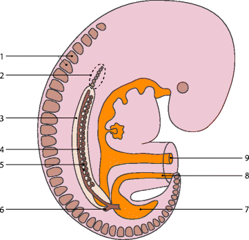
Fig. 1.2
1 Somites, 2 pronephros, 3 nephrogenic cord, 4 mesonephric duct, 5 mesonephric tubules, 6 metanephric diverticulum, 7 cloaca, 8 allantois, 9 omphaloenteric duct [Adapted from Moore [1]]
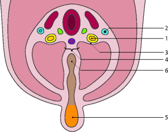
Fig. 1.3
1 Nephrogenic cord, 2 nephrogenic duct, 3 urogenital ridge, 4 dorsal mesentery, 5 umbilical vesicle, 6 midgut [Adapted from Moore [1]]
They appear late in the fourth week caudal to the rudimentary pronephroi.
They consist of glomeruli and tubules. The mesonephric tubules open into bilateral mesonephric ducts, which were originally the pronephric ducts.
The mesonephric ducts open into the cloaca.
The mesonephric ducts degenerate toward the end of the first trimester.
Metanephroi (Fig. 1.4)
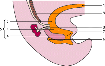
Fig. 1.4
1 Allantois, 2 vesical part, 3 pelvic part, 4 phallic part, 5 urogenital sinus, 6 rectum, 7 genital tubercle, 8 metanephric diverticulum, 9 mesonephros [Adapted from Moore [1]]
The primordial of permanent kidneys begins to develop early in the fifth week, from two different sources:
1.
The metanephric diverticulum is an outgrowth from the mesonephric duct near its intrance into the cloaca. The stalk of the diverticulum becomes the ureter and the cranial portion the collective system.
2.
The metanephrogenic blastema is derived from the caudal part of the nephrogenic cord.
1.1.1.2 Urinary Bladder
The expanded terminal part of the hindgut, the cloaca, is an endoderm-lined chamber that is in contact with the surface ectoderm at the cloacal membrane. The cloaca receives the allantois ventrally, which is a fingerlike diverticulum.
The cloaca is divided by a wedge of mesenchyme, the urorectal septum (Figs. 1.4 and 1.5), that develops between the allantois and the cloaca, in two parts:
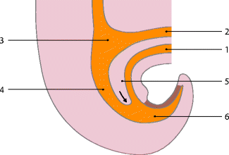

Fig. 1.5
1 Allantois, 2 omphaloenteric duct, 3 midgut, 4 hindgut, 5 urorectal septum (with arrow), 6 cloaca [Adapted from Moore [1]]
The rectum and cranial part of the anal canal dorsally
The urogenital sinus ventrally
By the seventh week, the urorectal septum has fused with the cloacal membrane (Fig. 1.6), dividing it into a dorsal membrane and a larger ventral urogenital membrane. The area of fusion of the urorectal septum with the cloacal membrane is represented in the adult by the perineal body.
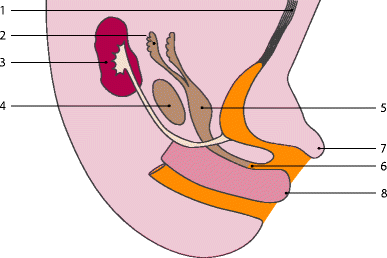
Fig. 1.6
1 Urachus, 2 uterine tube, 3 kidney, 4 ovary, 5 uterus, 6 vagina, 7 clitoris, 8 urorectal septum [Adapted from Moore [1]]
The urogenital system is divided in three parts:
A cranial vesical part that forms the bladder, but its trigone is derived from the caudal ends of the mesonephric ducts.
A middle pelvic part that becomes the entire urethra in females.
A caudal phallic part that grows toward the genital tubercule (primordium of the clitoris).
1.1.1.3 Urethra
The entire urethra is derived from endoderm of the urogenital sinus.
1.1.2 Development of the Gonads
The gonads are derived from three sources:
1.
Mesothelium (mesodermal epithelium) lining the posterior abdominal wall
2.
Underlying mesenchyme (embryonic connective tissue)
3.
Primordial germ cells
1.1.2.1 Indifferent Gonad
At the fifth week, a thickened area of mesothelium develops on the medial side of the mesonephros. Proliferation of this epithelium and the underlying mesenchyme produces a bulge on the medial side of the mesonephros—the gonadal ridge (Fig. 1.7). Fingerlike epithelial cords—the gonadal cords—soon grow into the underlying mesenchyme. The indifferent gonad consists of an external cortex and an internal medulla.
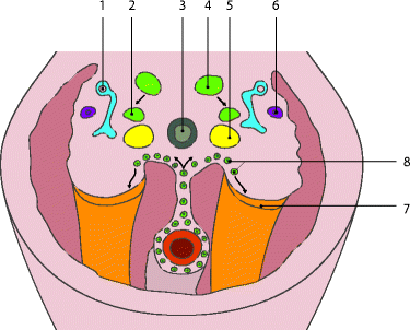

Fig. 1.7
1 Mesonephric duct, 2 suprarenal medulla, 3 aorta, 4 sympathic ganglion, 5 suprarenal cortex, 6 paramesonephric duct, 7 gonadal ridge, 8 primordial germ cells [Adapted from Moore [1]]
1.1.2.2 Primordial Germ Cells
These large spherical cells are visible early in the fourth week among the endodermal cells of the umbilical vesicle (yolk sac) near the origin of the allantois.
During folding of the embryo, the dorsal part of the umbilical vesicle is incorporated into the embryo.
As this occurs, the primordial germ cells migrate along the dorsal mesentery of the hindgut to the gonadal ridges (Fig. 1.8).
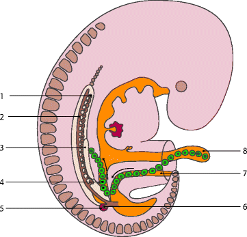

Fig. 1.8
1 Mesonephros, 2 mesonephric duct, 3 primordium of gonad, 4 primordial germ cells, 5 metanephrogenic blastema, 6 metanephric diverticulum, 7 allantois, 8 umbilical vesicle (yolk sac) [Adapted from Moore [1]]
During the sixth week, the primordial germ cells enter the underlying mesenchyme and are incorporated in the gonadal cords.
1.1.2.3 Sex Determination
Development of the male phenotype requires a Y chromosome. The SRY gene for a testis-determining factor (TDF) has been localized in the sex-determining short arm region of the Y chromosome. It is the TDF regulated by the Y chromosome that determines testicular differentiation. Under the influence of this organizing factor, the gonadal cords differentiate into seminiferous cords.
Development of the female phenotype requires two X chromosomes. A number of genes and regions of the X chromosome have special roles in sex determination. Consequently, the type of sex chromosome complex established at fertilization determines the type of gonad that differentiates from the indifferent gonad. The type of gonad present then determines the type of sexual differentiation that occurs in genital ducts and external genitalia.
Testosterone, produced by the fetal testes, dihydrotestosterone (see Chap. 3), a metabolite of the testosterone, and the anti-Mullerian hormone (AMH) determine normal male sexual differentiation. Primary female sexual differentiation in the fetus does not depend on hormones; it occurs even if the ovaries are absent and apparently is not under hormonal influence.
1.1.2.4 Development of Testes
TDF induces the gonadal cords to condense and extend into the medulla of the indifferent gonad, where they branch and anastomose to form the rete testis.
The rete testis becomes continuous with 15–20 mesonephric ducts that become efferent ductules. These ductules are connected with the mesonephric duct, which becomes the duct of the epididymis.
1.1.2.5 Development of the Ovaries
Before birth, the ovary is not identifiable histologically until approximately the tenth week.
Gonadal cords do not become prominent, but they extend into the medulla and form a rudimentary rete ovarii.
Cortical cords extend from the surface epithelium of the developing ovary into the underlying mesenchyme during the early fetal period (Fig. 1.9). This epithelium is derived from the mesothelium.
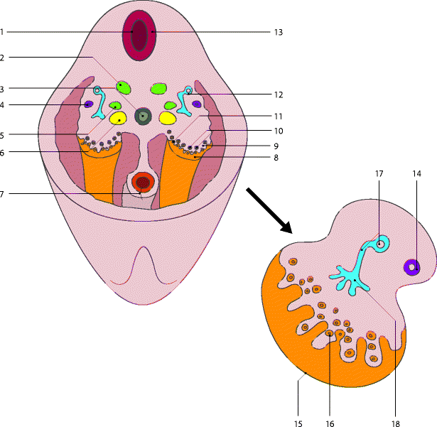

Fig. 1.9
(a) (At the top) 1 Neural tube, 2 sympathetic ganglion, 3 aorta, 4 paramesonephric duct, 5 primordium of suprarenal medulla, 6 primordium of suprarenal cortex, 7 hindgut, 8 gonadal ridge, 9 indifferent gona, 10 primordial germ cells, 11 primary sex cord, 12 mesonephric duct, 13 aggregation of neural crest cells. (b) (Below) 14 Uterine tube, 15 surface epithelium, 16 primordial, ovarian follicle, 17 mesonephric duct and tubule, 18 degenerating rete ovarii [Adapted from Moore [1]]
As the cortical cords increase in size, primordial germ cells are incorporated into them.
At 16 weeks, these cords break up into isolated clusters of primordial follicles each of which consists of an oogonium, derived from a primordial germ cell, surrounded by a single layer of flattened follicular cells derived from the surface epithelium.
Although many oogonia degenerate before birth, the two million or so that remain enlarge to become primary oocytes before birth.
After birth, the surface epithelium of the ovary flattens to a single layer of cells continuous with the mesothelium of the peritoneum at the hilum of the ovary.
1.1.3 Development of Genital Tract [1]
1.1.3.1 Development of the Female Genital Ducts and Glands
1.
During the fifth and sixth weeks, the genital system is in an indifferent state. Two pairs of genital ducts are present the mesonephric ducts (or Wolffian ducts) and paramesonephric ducts (or Muller ducts) (Fig. 1.7).
2.
The paramesonephric ducts develop lateral to the gonads and to mesonephric ducts on each side from longitudinal invaginations of the mesothelium on the lateral aspects of the mesonephroi. The edges of these paramesonephric grooves approach each other and fuse to form the paramesonephric ducts. The funnel shaped cranial ends of these ducts open into the peritoneal cavity. Caudally, the paramesonephric ducts run parallel to the mesonephric ducts until they reach the future pelvic region of the embryo. Here, they cross ventral the mesonephric ducts, approach each other in the median plane, and fuse to form a Y-shaped uterovaginal primordium (Fig. 1.6).
3.
This tubular structure projects in the dorsal wall of the urogenital sinus 1 (see definition later) and produces an elevation, the sinus tubercle, which defines the site of the future hymeneal membrane.
4.
Contact of the uterovaginal primordium with the urogenital sinus induces the formation of paired endodermal outgrowths—the sinovaginal bulbs. They extend from the urogenital sinus to the caudal end of the uterovaginal primordium. The sinovaginal bulbs fuse to form a vaginal plate. Later, the central cells of this plate break down, forming the lumen of the vagina. The epithelium of the vagina is derived from the peripheral cells of the vaginal plate.
Until fetal life, the lumen of the vagina is separated from the cavity of the urogenital sinus by a membrane—the hymen. The membrane is formed by invagination of the posterior wall of the urogenital sinus, resulting from expansion of the caudal end of the vagina. The hymen usually ruptures during the perinatal period.
The unfused portion of the ducts gives rise to the tubes. The uterovaginal primordium gives rise to the body and cervix of the uterus and superior part of the vagina.
The anatomical origin of tubes, uterus, and vagina is summarized in Table 1.1.
Table 1.1
Anatomical origin of tubes, uterus, and vagina
Muller ducts |
