Clinical indication
Examination
Radiopharmaceuticals
Typical time to completion
Sensitivity
Specificity
Positive predictive value
Negative predictive value
References
Acute pulmonary embolism
V/Q lung scintigraphy
Ventilation: 133Xe gas, inhaled, or 99mTc DTPA aerosol, inhaled
30–60 min
70–85 % for high-probability interpretation
96–98 % for normal/very low-probability interpretation
96–99 % for high-probability interpretation with high clinical suspicion
97–98 % for normal/very low-probability interpretation
Perfusion: 99mTc MAA, IV
Acute coronary syndrome
Myocardial perfusion imaging
99mTc sestamibi IV or 99mTc tetrofosmin IV
1–2 h
90–100 %
63–71 %
15–22 %
97–99 %
Acute biliary conditions: cystic duct obstruction (acute cholecystitis) and common bile duct obstruction
Hepatobiliary scintigraphy
99mTc IDA analog, IV
1–4 h
88–100 %
93–100 %
85–90 %
95–99 %
Acute biliary conditions: bile duct injury
1–2 h
100 %
90–100 %
91–100 %
100 %
[12]
Gastrointestinal bleeding
GI bleeding scintigraphy
99mTc RBCs, IV
1–2 h
78–97 %
70–100 % for diagnosis, 88–97 % for localization
75–77 %
76–93 %
Acute Pulmonary Embolism: Ventilation/Perfusion (V/Q) Lung Scintigraphy
Pulmonary embolism (PE) is a potentially fatal complication of deep venous thrombosis. In acute PE, thrombus dislodges from the vein, migrates to the pulmonary vasculature, and lodges in a main pulmonary artery or segmental branch. Thromboemboli reduce the cross-sectional area of the pulmonary arterial vascular bed, thus potentially resulting in hypoxia and hemodynamic compromise. Diagnosing acute PE can be very difficult clinically due to nonspecific symptoms and confounding clinical presentations mimicking other acute thoracic and abdominal conditions.
Both CT angiography (CTA) and ventilation/perfusion (V/Q) lung scintigraphy are well-accepted modalities for the imaging evaluation of suspected acute PE. Nowadays, CTA is performed in the vast majority of patients. However, V/Q scintigraphy remains relevant because radionuclide imaging of the lungs provides physiological information regarding not only regional pulmonary arterial perfusion but also bronchoalveolar ventilation. V/Q scintigraphy also spares the patient exposure to potentially nephrotoxic iodinated contrast and results in lower radiation dosimetry [3].
Although V/Q lung scintigraphy protocols vary by institution, ventilation images, using one of two commercially available radiopharmaceuticals, are typically acquired first. The perfusion phase using a standard radiopharmaceutical follows (Table 14.2). Perfusion imaging is based on the principle of capillary blockade. The radioactive MAA particles are larger than the capillaries and lodge in the precapillary arterioles; thus, their biodistribution reflects pulmonary arterial blood flow to both lungs. On these scintigraphic images, pulmonary segments with normal perfusion demonstrate uniform perfusion throughout (Fig. 14.1), while those with decreased perfusion demonstrate lower radioactivity than normal segments (Figs. 14.2 and 14.3). Classically, underlying lung disease presents as mildly to markedly abnormal perfusion that is “matched” by abnormal ventilation (Fig. 14.2). Acute PE affects perfusion only while ventilation should be preserved, resulting in the so-called V/Q mismatch pattern (Fig. 14.3). The PIOPED II criteria can be used to interpret V/Q lung scintigraphy (Table 14.3) [2].
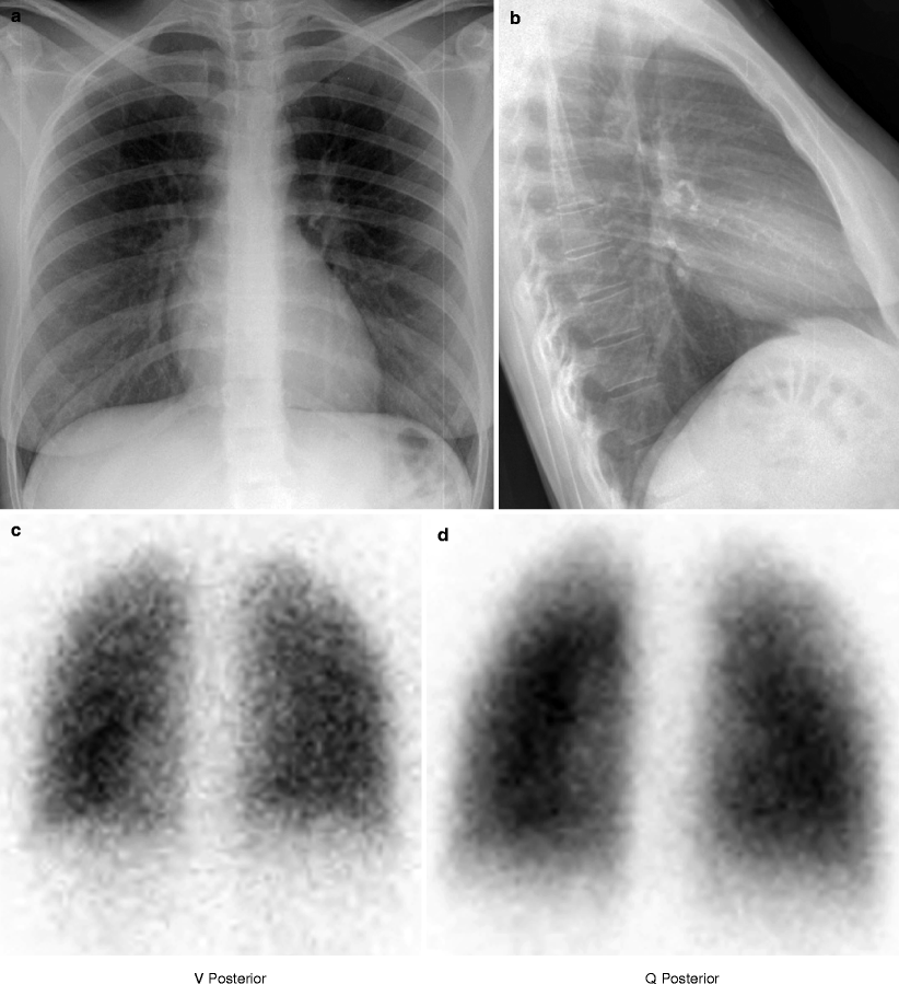
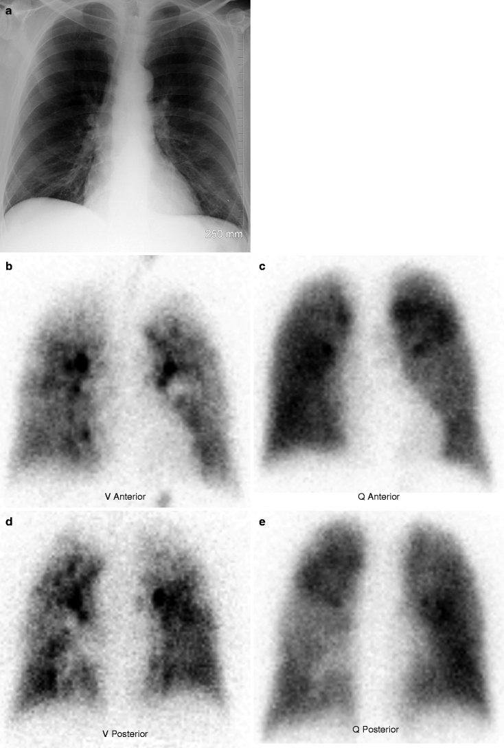
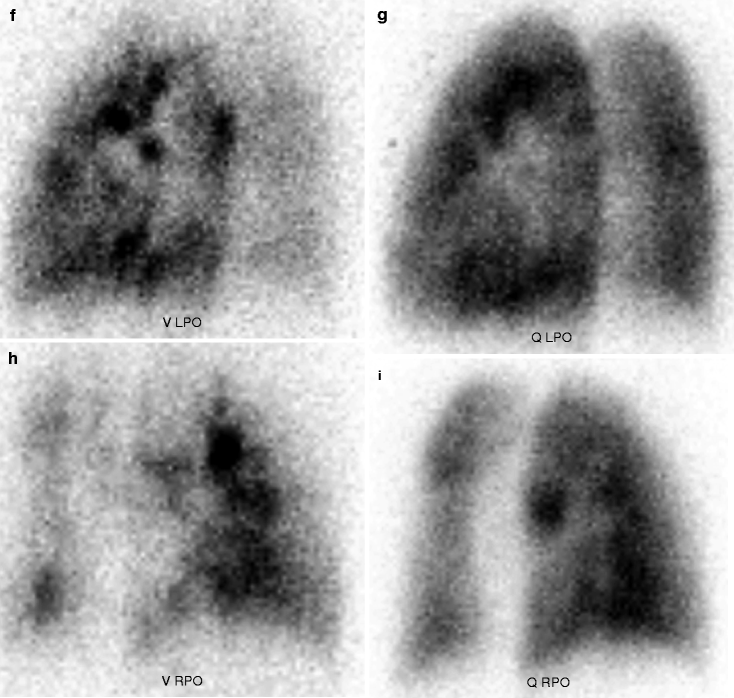
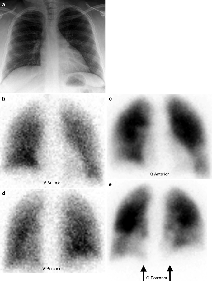
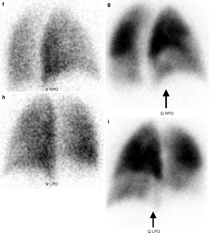
Table 14.2
Typical ventilation/perfusion (V/Q) lung scintigraphy technical protocol
1. Obtain contemporaneous chest radiography in posterior-anterior (PA) and lateral projections |
2. Patient inhales 20 mCi (740 MBq) of 133Xe gas or 3 mCi (111 MBq) of aerosolized 99mTc diethylenetriaminepentaacetic acid (DTPA) |
3. Acquire ventilation images in posterior (133Xe gas) or anterior, posterior, bilateral anterior oblique, and bilateral posterior oblique (99mTc DTPA aerosol) projections |
4. Administer 4 mCi (148 MBq) of 99mTc macroaggregated albumin (MAA) IV |
5. Acquire perfusion images in anterior, posterior, bilateral anterior oblique, and bilateral posterior oblique projections |
Fig. 14.1
Normal. Normal posterior-anterior (a) and lateral (b) chest radiographs. Normal ventilation (c) and perfusion (d) scintigraphy performed with 133 Xe gas (ventilation) and 99mTc MAA (perfusion)
Fig. 14.2
Multiple matched V/Q abnormalities related to underlying airways disease. Normal chest radiograph (a). Heterogeneous 99mTc DTPA ventilation (left panel: b, d, f, h) and heterogeneous 99mTc MAA perfusion (right panel: c, e, g, i)
Fig. 14.3
High probability for acute PE (multiple V/Q mismatches). Normal chest radiograph (a). Normal 99mTc DTPA ventilation (left panel: b, d, f, h) and abnormal 99mTc MAA perfusion (right panel: c, e, g, i) with multiple bilateral lower lobe segmental perfusion defects (arrows)
Table 14.3
Simplified criteria for interpretation of V/Q lung scintigraphy for diagnosis of acute PE
Interpretation | Patterns on V/Q lung scintigraphy |
|---|---|
PE absent: “normal” | No perfusion defects |
PE absent: “very low probability” | Nonsegmental perfusion defects (e.g., cardiomegaly, enlarged hila, elevated hemidiaphragm) |
Perfusion defect smaller than corresponding X-ray abnormality | |
Two or more V/Q matches with corresponding normal chest X-ray and otherwise relatively normal perfusion | |
Three or fewer small segmental perfusion defects | |
Triple-matched V/Q/X-ray abnormality in upper/mid-lung | |
Stripe sign (i.e., preserved perfusion to pleural surface) | |
Large pleural effusion with otherwise normal perfusion | |
Nondiagnostic for PE: “low probability” or “intermediate probability” | All other patterns, including triple-matched V/Q/X-ray abnormality in lower lung |
PE present: “high probability” | Two or more segmental V/Q mismatches |
In selecting the optimal radiological approach for suspected acute PE, the emergency physician should take into account multiple factors, not the least of which include sensitivity and specificity of the different imaging examinations, technical availability, and patient safety. First, for a patient with acute symptomatology, conventional chest radiography remains first-line. Patients with significantly abnormal chest radiographs should be directed to CTA and are not well suited for V/Q lung scintigraphy because there is greater likelihood of a nondiagnostic interpretation due to confounding underlying pulmonary conditions such as chronic obstructive lung disease, pneumonia, pleural effusion, or atelectasis [17]. Concomitantly, there can be considerable interobserver variability of V/Q interpretations, especially in low and intermediate categories. CTA has the distinct advantage of evaluating the entire chest and upper abdomen and can delineate alternate thoracic or abdominal pathologies.
Second, in most institutions today, CTA is available around the clock with an on-site CT technologist who can perform the examination in a timely manner. During the standard workday, V/Q lung scintigraphy can be performed relatively quickly; however, during the off-hours, the NM technologist usually needs to travel into the hospital, and the radiopharmaceuticals may need to be prepared in-house or delivered from an outside pharmacy, all requiring additional time.
Third, there is much concern about radiation dosimetry, and the risks related to iodinated contrast are well established [3]. With CTA, there is higher radiation exposure to the patient, particularly to the female breast. The tissue dose to the breasts of nonpregnant women can be 10–60 mGy (1–6 rad) and is probably higher in pregnancy. Conversely, the breast dose from V/Q lung scintigraphy can be much lower than 0.31 mGy (0.031 rad) or almost 200 times less than CTA, and during the first trimester of pregnancy, the fetal dose can be halved using a modified reduced dose V/Q lung scintigraphy protocol [3]. Thus, for selected patients with special medical considerations such as pregnancy, breast-feeding, poor renal function, or IV contrast allergy, V/Q lung scintigraphy may represent the most appropriate imaging examination.
In summary, V/Q lung scintigraphy can be considered as a primary imaging approach in patients with suspected acute PE when there is:
1.
Normal chest radiography
2.
No concurrent cardiopulmonary process
3.
Available NM facilities and personnel
4.
Relative contraindication to CTA regarding radiation exposure or use of iodinated contrast
Acute Coronary Syndrome: Myocardial Perfusion Imaging
Acute coronary syndrome (ACS) accounts for approximately 10 % of all emergency department visits, making it one of the most commonly encountered medical emergencies. ACS refers to a spectrum of clinical presentations ranging from ST-segment elevation myocardial infarction to unstable angina. Clinical presentation, electrocardiography (ECG), and cardiac biomarkers, such as troponin, guide initial risk stratification. Troponin has become the favored biomarker for determination of myocardial necrosis because of the high sensitivity and specificity. However, myocardial biomarkers can only diagnose infarction and cannot identify ischemia in the absence of necrosis; also, laboratory evidence lags behind the physiological event. By convention, patients are stratified into three risk groups: high risk, moderate risk, and low risk.
The majority of patients with chest pain have no history of coronary artery disease and no ischemic ECG changes. Risk of ACS in such patients is low; however, it is not zero. Identification of high-risk patients within this cohort can be difficult clinically. Thus, many patients without ischemia are admitted for observation and further testing. Conversely, despite a low threshold for admission, a significant minority of patients with atypical symptoms of ACS who actually have acute myocardial infarction are initially triaged as low risk and are unintentionally discharged home [18].
Myocardial perfusion imaging (MPI) can be effectively used as a triage tool in patients with chest pain of unclear etiology. MPI typically utilizes one of two available 99mTc radiopharmaceuticals, 99mTc sestamibi and 99mTc tetrofosmin (Table 14.4). The biodistribution within the left ventricular myocardium is generally proportional to coronary arterial blood flow at the time of IV administration; these tracers do not redistribute for several hours. Normally, there is uniform myocardial perfusion (Fig. 14.4). If a patient with ACS is injected at rest while experiencing coronary-related chest pain, the distribution of the perfusion agent will be altered and will demonstrate diminished regional perfusion corresponding to the vascular territory involved (Fig. 14.5). However, acute or prior myocardial infarction may produce a similar perfusion defect on rest MPI; therefore, differentiation between ischemia and infarction is not possible with rest MPI alone.
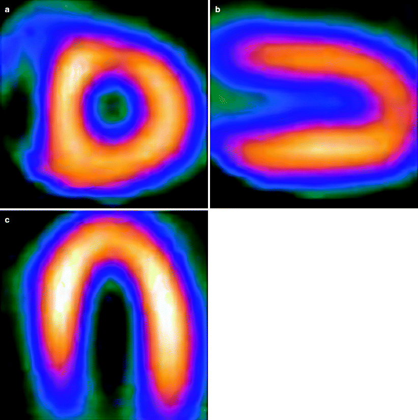
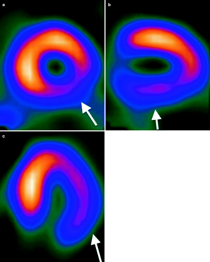
Table 14.4
Typical rest with chest pain myocardial perfusion imaging (MPI) technical protocol
1. Administer 25 mCi (925 MBq) of 99mTc sestamibi or 99mTc tetrofosmin IV while patient has chest pain at rest |
2. Wait 15–30 min |
3. Perform single-photon emission computed tomography (SPECT) imaging |
4. Evaluate rest-only images |
Fig. 14.4
Normal rest MPI. Uniform 99mTc sestamibi activity throughout the left ventricular myocardium. (a) Short-axis, (b) vertical long-axis, and (c) horizontal long-axis images
Fig. 14.5
Abnormal rest MPI. High-grade occlusion of left circumflex coronary artery as etiology of active chest pain during 99mTc sestamibi administration IV. Large, moderately severe perfusion defect in the inferior-lateral wall extending from apex to base (arrows). (a) Short-axis, (b) vertical long-axis, and (c) horizontal long-axis images
Rest MPI in symptomatic patients has reported sensitivities of 90–100 % with negative predictive values (NPV) of greater than 99 % for identifying patients without cardiac events [19]. The high sensitivity of rest MPI is dependent on the presence of chest pain during tracer injection; that is, MPI in patients injected after cessation of chest pain does not yield the same sensitivity, and that subgroup should undergo stress testing. Thus, the high NPV of rest MPI in patients with chest pain allows the emergency physician to establish confidently the absence of myocardial ischemia or infarction as the etiology of the symptomatology. Specifically, negative rest MPI directs disposition of patients who might otherwise have a prolonged hospital stay, and, conversely, positive rest MPI identifies high-risk patients who might be categorized incorrectly as low risk and might have a delayed diagnosis of ACS.
Incorporation of MPI into the acute chest pain diagnostic algorithm is beneficial. The ERASE trial [20] demonstrated that MPI in the emergency setting reduced unnecessary admissions without increasing inappropriate discharges. MPI as a triage tool is most effective when used on low-risk patients who are experiencing active chest pain (i.e., hemodynamically stable, no ECG changes, no prior coronary disease). Together, concordantly negative rest MPI and negative biomarkers identify patients who can safely be discharged, or, alternatively, undergo early stress testing with or without hospital admission depending on those results. MPI is the only commonly available imaging technique that provides a direct and accurate assessment of myocardium at risk [19]. The greatest benefit of MPI lies in its high NPV, which assists the emergency physician in effectively excluding ACS in low-risk patients, lowering costs, shortening length of hospital stay, and decreasing morbidity [7, 21, 22].
Related posts:


Stay updated, free articles. Join our Telegram channel

Full access? Get Clinical Tree


