10 Evaluation of Collateral Ligaments
Ulnar Collateral Ligament
♦ Setup
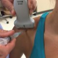
• The patient is seated in a chair with the arm placed on a table between the operator and the patient (Fig. 10.1).
• The table should be draped with a towel or sheets to increase patient comfort.
• The elbow is mildly flexed and the upper arm is externally rotated to expose the medial aspect of the elbow.
• A high-frequency (12-15 MHz) linear transducer is used for the examination. A high-frequency small-footprint transducer (hockey stick) can be used as needed.

Fig. 10.1 Patient setup for an ulnar collateral ligament evaluation. The patient’s arm is placed on the examination table with the elbow mildly flexed and the upper arm externally rotated to expose the medial elbow.
♦ Landmarks
The medial epicondyle should be located (Fig. 10.2).
♦ Probe Positioning
• The medial epicondyle should be identified with palpation.
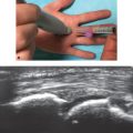
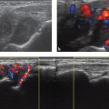
• The proximal portion of the transducer should be placed on the medial epicondyle (Fig. 10.3a).
• With the proximal portion of the transducer anchored on the medial epicondyle the distal aspect of the transducer is rotated until it is parallel with the long axis of the forearm.
• Without rotating the transducer, it is moved in the dorsal-ventral plane until the anterior bundle of the ulnar collateral ligament is visualized.
• The entire ligament should be visualized from the humeral attachment to the ulnar attachment in a single image (Fig. 10.3b).
• The ligament is relatively hyperechoic with a fibrillar pattern.
• The common flexor-pronator tendon can be visualized superficial to the ulnar collateral ligament.
• Transverse images can also be acquired as needed.
• Fig. 10.3b,c demonstrates the differences in resolution between a linear 15 MHz transducer and a linear hockey stick 18 MHz transducer. Increased resolution is observed with the 18 MHz hockey stick transducer at the expense of a smaller field of view and reduced tissue penetration.

Fig. 10.2 Surface landmark for ulnar collateral ligament evaluation; the yellow circle marks the medial epicondyle.

♦ Normal Anatomy
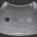
• The ulnar collateral ligament is composed of three bands: anterior, posterior, and transverse (oblique).
• The anterior band, extending from the medial epicondyle to the sublime tubercle of the ulna, plays the most important role in resisting valgus stress to the elbow (Fig. 10.4).
• The proximal insertion can be broad and fanlike with a prominent fibrillar pattern. This appearance should not be confused with pathology.
• The distal attachment on the sublime tubercle can be seen up to 3 mm distal to the articular margin. More distal insertions should be interpreted as pathologic.
♦ Pathology
• Partial thickness tears are observed as areas of heterogeneous echotexture with loss of the normal fibrillar pattern. Disruption of portions of the ligament and areas of hypoechogenicity can also be observed.
• Full-thickness tears are characterized by complete gaps in the ligament filled with isoechoic signal (fluid or hemorrhage).
• Chronic injuries can cause ligament laxity without disruption.

Fig. 10.4 Ulnar collateral ligament.
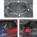
• Dynamic imaging can be used to increase confidence in the degree of injury.
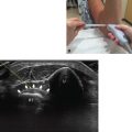
○ The elbow is flexed to approximately 30 degrees, and valgus stress is placed on the elbow. An assistant can be used to apply valgus stress while the ultrasonographer controls the transducer (Fig. 10.5 and Video 10.1).
○ Images of the medial ulnohumeral joint and ulnar collateral ligament are obtained with and without stress.
○ With full-thickness tears, separation of the fibers can be seen.
○ The joint space is also measured with and without stress. Joint space widening greater than 1 mm is concerning for pathology.
○ Comparison to the contralateral side is then performed.
○ Notably, asymptomatic professional baseball pitchers have demonstrated asymmetric increased joint space widening in their pitching arms compared with their nondominant arms. Widening of the dominant arm may be greater than 1 mm.