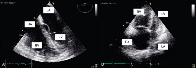
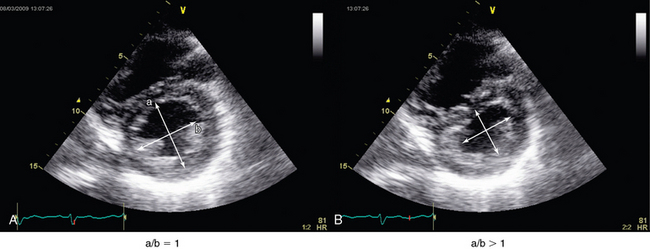
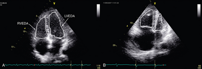
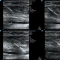
33 Assessment of right ventricular (RV) function is a key element in the hemodynamic evaluation of patients in the intensive care unit (ICU) with shock or respiratory failure.1,2 For several decades the former was considered to play a minor role in hemodynamic failure. However, RV dysfunction may be a crucial element in the hemodynamic compromise that intensivists encounter in various clinical scenarios such as pulmonary embolism (PE), sepsis and septic shock, myocardial infarction, and acute respiratory distress syndrome (ARDS).3–5 The right ventricle is highly sensitive to changes in preload and afterload and, unlike the left ventricle (LV), may become acutely enlarged. A pulmonary artery catheter can be used to estimate right atrial pressure (RAP), pulmonary arterial pressure (PAP) and resistance, and cardiac output. In recent years, echocardiography has progressively replaced the former in the ICU. Echocardiography facilitates accurate, noninvasive, prompt, and serial evaluation of RV function in ICU patients. The configuration of the right ventricle is rather complex in that it has two perpendicular chambers—a horizontal one corresponding to the filling chamber (the sinus) and a vertical one corresponding to the outflow tract (the cone or infundibulum). The right ventricle has a normal free wall thickness of 4 mm and is less muscular than the LV since it is adapted to the pulmonary vasculature. It is very sensitive to changes in afterload and becomes acutely dilated; its ejection fraction is radically decreased whenever abrupt increments in PAP occur. In the latter case, RV pressure may become transiently greater than pressure in the LV and thus cause end-systolic bowing of the interventricular septum into the LV.6,7 Chronically increased PAP induces RV hypertrophy. Because the heart is enveloped by the pericardium and the right ventricle is wrapped around the LV, any modification in one ventricle will modify the size and the function of the other (left-right ventricular interdependency). The LV normally contributes 25% of RV contraction, which may be increased up to 35% in patients with pulmonary hypertension.7,8 The right ventricle is very sensitive to coronary flow and is perfused, unlike the LV, during both systole and diastole.9 RV size may be assessed by measurement of volume. Two-dimensional (2D) echocardiography is unable to provide an accurate estimate of RV volume because of its complex shape. Three-dimensional (3D) echocardiography is more accurate. However, it is time-consuming and not yet readily available, and a perfect view of the endocardial contour is required (which is hard to achieve in the critically ill). Vieillard Baron et al proposed measuring the right (RVEDA) and left (LVEDA) ventricular end-diastolic areas by tracing the endocardium on an apical four-chamber view.10 When the endocardium is not well identified, the area should be traced to the epicardium. The RVEDA/LVEDA ratio was assessed in normal subjects and found to vary between 0.36 and 0.60. Thus a ratio lower than 0.6 (Table 33-1) is considered normal, whereas ratios between 0.6 and 1 and higher than 1 correspond to moderate and severe RV dilatation, respectively (Figures 33-1 and 33-2). This sonographic evaluation is performed by using either transthoracic (TTE) or transesophageal (TEE) echocardiography.10 The American Society of Echocardiography and the European Association of Echocardiography have published guidelines for evaluation of RV function by echocardiography11; Howeverhowever, several of the recommendations are not easily applicable in the ICU. TABLE 33-1 Evaluation of Right Ventricular Function by Echocardiography: Abnormal Values of Various Doppler and Two-Dimensional Echocardiographic Indices Data from Jardin F, Dubourg O, Bourdarias JP: Echocardiographic pattern of acute cor pulmonale, Chest 111:209-217, 1997; and Rudski LG, Lai WW, Afilalo J, et al: Guidelines for the echocardiographic assessment of the right heart in adults: a report from the American Society of Echocardiography endorsed by the European Association of Echocardiography, a registered branch of the European Society of Cardiology, and the Canadian Society of Echocardiography, J Am Soc Echocardiogr 23:685-713, quiz 786-788, 2010. Figure 33-2 Transthoracic echocardiographic apical four-chamber view: assessment of the ratio of right ventricular to left ventricular end-diastolic area in a normal patient (A) and one with acute cor pulmonale (B). Alterations in RV shape may be used to assess dilatation. As it dilates, the right ventricle loses its characteristic triangular shape and becomes more rounded. Quantitative measurement of RV size by means of the RVEDA/LVEDA ratio is simple and has good reproducibility. Charon et al proposed using subjective (qualitative) assessment of RV size by eyeballing the right ventricle on an apical four-chamber view since they found significant correlation with measurements of the RVEDA/LVEDA ratio.12 The subcostal view, which is usually more accessible than other views (e.g., apical) in mechanically ventilated patients, may be used to perform RV measurements in the ICU. The interventricular septum should be analyzed in patients with RV dysfunction. It is best analyzed on a parasternal short-axis view at the level of left ventricular papillary muscles. In normal subjects, left ventricular pressure is always higher than RV pressure. Consequently, on a parasternal short-axis view the septum bows into the right ventricle, thus giving the LV its circular shape (O shape). Whenever RV systolic overload occurs, RV end-systolic pressure exceeds that in the LV (because of a prolongation in RV contraction), and this causes the interventricular septum to move toward the LV (septal dyskinesia). This flattening of the septum produces a D-shaped LV and represents a qualitative assessment of this paradoxic septal movement. In addition, a quantitative evaluation can be achieved by direct measurement of the systolic eccentricity index.10–12 The latter, which is measured on a parasternal short-axis view at the level of left ventricular papillary muscles, is the ratio of the diameter that bisects the papillary muscle to the perpendicular diameter at end-systole. In septal dyskinesia, the systolic eccentricity index is higher than 1 (Figure 33-3; also see Figure 33-2).
Evaluation of right ventricular function in the intensive care unit by echocardiography
(CONSULTANT-LEVEL EXAMINATION)
Overview
Right ventricular physiology
Evaluation of right ventricular function by echocardiography
Right ventricular size
Abnormal Value
Maximal RV diameter (parasternal view) (see Figure 33-3)
>42 mm
RVEDA/LVEDA (apical 4-chamber view) (see Figures 33-1 and 33-2)
>0.6
RV two-dimensional fractional area contraction (apical four-chambers view)
<35%
RV ejection fraction
<44%
RV free wall thickness
>4 mm
SPAP (on tricuspid regurgitation or pulmonary regurgitation; see Figures 33-5 and 33-6C)
>35-40 mm Hg
TAPSE (see Figure 33-4A)
<16 mm
S on TDI (see Figure 33-4B)
<0 cm/sec
Interventricular septal shape and movement
Assessment of right ventricular systolic function
Right ventricular fractional area change
Related posts:
 Ultrasound-guided peripheral intravenous access
 Ultrasound-guided arterial catheterization
 Ultrasound-guided placement of inferior vena cava filters: (CONSULTANT-LEVEL EXAMINATION)
 Use of ultrasound in the evaluation and treatment of intraabdominal hypertension and abdominal compartment syndrome
 Ultrasound imaging in space flight
 Procedural ultrasound for surgeons: (CONSULTANT-LEVEL EXAMINATION)
![]()
Stay updated, free articles. Join our Telegram channel

Full access? Get Clinical Tree


Evaluation of right ventricular function in the intensive care unit by echocardiography: (CONSULTANT-LEVEL EXAMINATION)