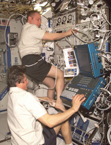
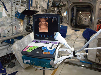
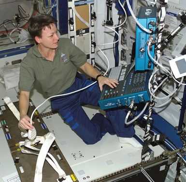
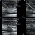
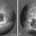
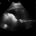
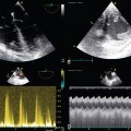
50 Between the 1970s and mid-1990s, eight different ultrasound imagers were flown on National Aeronautics and Space Administration (NASA) and Russian spacecraft and successfully operated for research purposes. These successes justified the installation of the first permanent ultrasound system (HDI-5000, ATL/Philips, Andover, MA) for the ISS Human Research Facility (2002). After serving for 10 years, the ISS Ultrasound-1was replaced by a new unit with advanced research and clinical capabilities (Figure 50-1). Figure 50-1 The International Space Station (ISS) Ultrasound System (Vivid Q, General Electric, Waukesha, WI) is ready for use. The box underneath the machine is a data and power interface. Note the oversized water drop on the 12-MHz linear array transducer, to be used for acoustical coupling instead of gel. Also note the multicolor keyboard overlay specially designed for remote guidance purposes. (Image courtesy National Aeronautics and Space Administration.) Significant efforts were needed before space-based ultrasound imaging could be recognized for clinical use, with no medical expertise onboard and little evidence on the imaging representations of disease in microgravity.1 The successful implementation of clinical ultrasound in space flight is a great testament to the essential universality and versatility of ultrasound imaging. We hope this chapter will motivate more physicians to seek and promote imaging solutions in their areas of practice. Space medicine depends on ultrasound imaging more than most other clinical disciplines because of the absence of other diagnostic imaging modalities and the operational nature of the setting with limited resources and the very limited ability to safely and quickly evacuate the ill or injured crew member. Depending on the clinical and operational circumstances, a focused diagnostic examination in space with a single binary clinical question (emergency medicine model) can evolve into a broader, multitarget assessment and monitoring (critical care model) or into a comprehensive and specific imaging application (radiology model). The current medical requirements for the International Space Station foresee a possibility of advanced life support of a seriously ill or injured crew member for up to 72 hours, which, regardless of the initial offending factors, essentially includes close monitoring of the hemodynamic parameters, pulmonary physiology, airway management, intracranial pressure, and so forth, without the trained personnel and resources taken for granted in any terrestrial hospital. Therefore space medicine experts have great interest in both established and emerging ultrasound applications, and they monitor the recently accelerating implementation of bedside ultrasound approaches and techniques in terrestrial emergency medicine and intensive care. In the absence of gravity, the patient must often be physically restrained by using elastic cords or fabric belts to ensure positional stability; in general, the mutual positioning of the operator, patient, and imaging hardware must be globally compatible with the medical equipment setup for emergency medical treatment and life support activities. Thus a seriously ill patient would be scanned on the special electrically isolated restraint system designed for advanced life support procedures (Figure 50-2). The operator should also be restrained in a comfortable and sustainable position to consistently exert a contact force on the transducer and have both hands available for the imaging procedure. Figure 50-2 Astronaut Peggy Whitson (International Space Station [ISS]-5 expedition, 2002) developing the optimal setup for ultrasound examinations on the ISS by using the Crew Medical Restraint System (CMRS). Note the use of foot restraints. This position was found unacceptable because applying force on the transducer would make the operator position unstable. The CMRS was moved closer and was used for restraining both the subject (using straps) and the operator (inserting one knee into the space under CMRS). (Image courtesy National Aeronautics and Space Administration.) Self-scanning is also possible with minimal foot restraint, except for patients in distress or when a more experienced operator is available. Furthermore, some crews use creative ways of positioning in microgravity for specific applications (Figure 50-3). In the lack of gravity, the position of an object or the shape and distribution of a fluid collection are determined by the combined effect of weaker physical forces, such as properties of the fluid; surface interaction forces; tissue and organ compliance; random pressure fluctuations and gradients, such as peristalsis; and small accelerations. The absence of gravity may thus require significant modifications of imaging techniques as well as data interpretation. A terrestrial “gold standard” imaging procedure may not work in microgravity, whereas a previously unexplored technique may be the method of choice. Animal models of internal bleeding2–4 in simulated microgravity strongly suggest high diagnostic accuracy of ultrasonography if performed and interpreted using microgravity-based evidence and considerations. Intrathoracic hemorrhage and pneumothorax, maxillary sinusitis, lung abscess, ileus with dilatated bowel loops, and urinary calculi are examples of the many imaging situations influenced by the gravity vector or its absence.
Ultrasound imaging in space flight
Scope of diagnostic ultrasound in space
Implications of microgravity
Patient and operator positioning
Normal and pathologic anatomy
Lack of imaging expertise onboard
Related posts:
 Ultrasound-guided peripheral intravenous access
 Ultrasound-guided arterial catheterization
 Ultrasound-guided placement of inferior vena cava filters: (CONSULTANT-LEVEL EXAMINATION)
 Use of ultrasound in the evaluation and treatment of intraabdominal hypertension and abdominal compartment syndrome
 Evaluation of right ventricular function in the intensive care unit by echocardiography: (CONSULTANT-LEVEL EXAMINATION)
 Procedural ultrasound for surgeons: (CONSULTANT-LEVEL EXAMINATION)
![]()
Stay updated, free articles. Join our Telegram channel

Full access? Get Clinical Tree


Ultrasound imaging in space flight