14 Ultrasound guidance to obtain vascular access has become increasingly more popular over the past decade. Advances in ultrasound technology have enhanced its ability to detect and interrogate smaller vascular structures for cannulation purposes. Given the potential complications that can develop when accessing central veins, practitioners have turned to ultrasound-guided peripheral access. Although no invasive procedure is without risks, the relative array of complications potentially produced by peripheral, non–centrally extending venous lines tend to be significantly less severe than those from central venous access.1 This chapter outlines the indications, risks, and benefits of ultrasound-guided, noncentral peripheral intravenous (IV) access. The standard limitations described with landmark-based IV catheter placement can be overcome given the advances in visualization brought to the bedside with ultrasound. With regard to operators, emergency physicians, anesthesiologists, nursing staff, intensivists, and their respective trainees have reported peripheral IV cannulation success rates of higher than 85%.2–7 With regard to risk for infection, Maki et al reported that peripheral IV catheters in general maintain lower infection rates than do any other forms of IV access.1 Additionally, when complications did occur, they tended to not be as morbid as those associated with central venous catheters. Despite such a successful track record of placement and safety, limited data exist on the role of ultrasound-guided peripheral IV access in patients in the intensive care unit (ICU). One of the reasons for this is that many medications should not be administered through peripheral access given their potentially damaging effects on veins (e.g., certain vasopressors, total parenteral nutrition [TPN]). In another report, medications such as vancomycin or phenytoin were suggested to be associated with increased rates of thrombophlebitis.8 Aside from these relative contraindications, a question remains: in patients who do not need central venous access, are ultrasound-guided peripheral IV techniques reliable enough to allow increased avoidance or removal of central lines in the critically ill? Gregg et al were able to achieve ultrasound-guided peripheral venous access in 99% of patients who failed standard landmark attempts at placement.7 Among the diverse group of trauma, postsurgical, and cardiac intensive care patients, 34 central lines were avoided and 40 were removed as a result of adequate peripheral access. In a small randomized controlled trial, Kerforne et al also demonstrated increased success rates of ultrasound-guided peripheral IV access over the landmark technique in critical care patients who were deemed to have difficult access.9 Despite reports of relatively low complication rates with ultrasound-guided peripheral IV access in the ICU,7 no trials have directly compared rates of infection or thrombophlebitis with the use of ultrasound-guided versus landmark techniques. Different techniques for placing peripheral IV catheters have been described. One-person operation of the ultrasound transducer while placing the IV line has the benefit of allowing the entire procedure to be performed by a single party. However, applying appropriate skin tension when limited to a single hand to perform the insertion has the potential to be awkward. This has led to reports of a two-person technique that involves an ultrasound operator and an IV operator working together throughout the placement procedure. Chinnock et al showed that the number of operators placing the IV line was not predictive of successful placement,10 and a variety of other studies describing either a one- or two-person technique have shown successful placement more than 85% of the time.2–7 Another variation in equipment described includes the use of guidewire assistance. Guidewire-assisted vascular access devices have traditionally been used when attempting arterial access; however, given their ease of use, successful single-operator cannulation rates higher than 95% can be achieved.7–11
Ultrasound-guided peripheral intravenous access
Related posts:
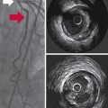 Intravascular ultrasound: (CONSULTANT-LEVEL EXAMINATION)
 Integrating picture archiving and communication systems and computerized provider order entry into the intensive care unit: The challenge of delivering health information technology–enabled innovation
 Endobronchial ultrasound: (CONSULTANT-LEVEL EXAMINATION)
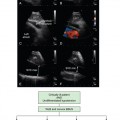
 Ultrasonography in circulatory failure
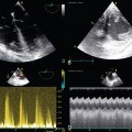 Evaluation of right ventricular function in the intensive care unit by echocardiography: (CONSULTANT-LEVEL EXAMINATION)
 Procedural ultrasound for surgeons: (CONSULTANT-LEVEL EXAMINATION)
![]()
Stay updated, free articles. Join our Telegram channel

Full access? Get Clinical Tree


