7 Evaluation of Wrist Joints
Ultrasonography is a suitable imaging modality to evaluate wrist joints. Ultrasound exploration of the wrist is very helpful in the detection of synovitis, crystal deposits, and bone changes such as erosions and osteophytes. Joint aspiration for diagnostic purposes and therapeutic guided injections of wrist joints are other elegant attributes of ultrasound.
♦ Setup
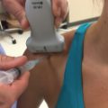
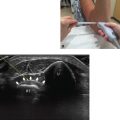
• The patient should be seated with the hand resting in neutral position with the palm down on the table for ultrasound examination of the dorsal aspect (Fig.7.1).
• For ultrasound examination of the ventral aspect, the hand should be resting in neutral position or with slight dorsal extension with the palm up. A rolling pad placed under the wrist may be helpful.
• A linear probe with high frequency (10-18 MHz) should be used. Small transducers (e.g., hockey stick) are more dependable for assessing multiplanar joints of the wrist.

Fig. 7.1 Positioning for ultrasonographic examination of the wrist. (a) Wrist in neutral position with the palm down. (b,c) Wrist in neutral position with the palm up and with minimal dorsal extension.
♦ Landmarks
• Lister’s tubercle is a bony prominence that can be palpated at the dorsal aspect on the distal extreme of the radius, at the same level of the ulnar styloid (Fig.7.2).
• The ulnar styloid is a bony prominence of the distal ulna, easily recognizable by inspection or palpation of the dorsal aspect of the forearm.
• The scaphoid (ventral aspect) is a lateral bony prominence at the base of the thumb. It can be palpated at the same level (distally to the joint line) of the pisiform bone, which is medially located.

Fig. 7.2 Useful bony landmarks of the wrist at the dorsal and ventral aspect. The prominence of the scaphoid bone (S) can be palpated in the area at the base of the thumb. LT, Lister’s tubercle; US, ulnar styloid.
♦ Probe Positioning
Axial
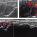
• The probe should be placed on the transverse plane at the proximal level of the joint line (Fig.7.3).
• The probe should be swept distally with the hand in neutral position, followed by a provocative stress test with ulnar deviation to evaluate the integrity of the scapholunate ligament or the presence of any abnormal gap between scaphoid and lunate bones.

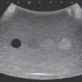
Fig. 7.3 Examination of the distal radioulnar and scapholunate joints. (a) The probe is oriented in the short axis at the joint line; then, using Lister’s tubercle (LT) as a landmark, the probe is moved distally. (b) Transverse ultrasound image showing the distal radioulnar joint and the radioulnar recess (asterisk). (c) Transverse view of the scaphoid and lunate bones. *, Scapholunate joint.
Longitudinal
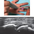
• The probe should be placed on Lister’s tubercle and moved slightly distally; it is then swept laterally and medially (Fig.7.4).
• The probe should then be placed on the ulnar styloid, oriented in a sagittal plane (Fig.7.5).
• A coronal plane is also helpful for assessing the ulnar bone and the triangular fibrocartilage complex.
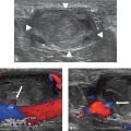
• The probe is next placed in a sagittal oblique plane on the base of the thumb (ventral aspect of the wrist). After the base of the first metacarpal bone is identified, the probe is moved proximally (Fig.7.6). Multiplanar assessment by ultrasonography can reveal degenerative bone changes that cannot be seen in a single radiographic plane.1
1Checa A, Panahi E. Clinical images: is ultrasonography better than radiography for determining the need for surgical treatment in patients with carpometacarpal osteoarthritis? Arthritis Rheum 2014;66(7):1954.