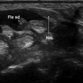
Fig. 3.1
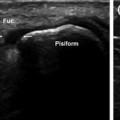
Tenosynovitis of the extensor brevis and abductor longus of the thumb (Est b—Abd l) caused by a radial fracture. During movement of the wrist a, b, c, the stump of the fractured bone (arrow) impinges on the sheath surrounding the abductor longus and extensor brevis tendons, and the repeated microtrauma leads to tenosynovitis. The multilamellar abductor longus represents an anatomic variant
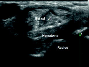
Fig. 3.2
Recent mid-epiphyseal fracture of the radius with multiple fragments, hematoma, and tenosynovitis of the extensor digitorum communis and extensor indicis proprius tendons (Est cd—pi). During wrist movement, the large, partially organized hematoma surrounding the fracture impinges on the sheath of the fourth compartment, causing friction and related inflammation
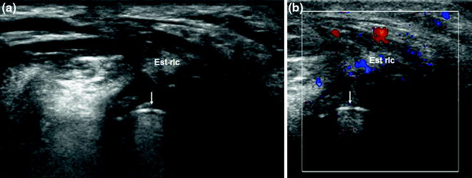
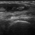
Fig. 3.3
Tenosynovitis of the extensor carpi radialis longus tendon related to orthopedic hardware used to set a fracture of the scaphoid bone. During wrist movement, metal screws used to achieve osteosynthesis (arrow)—represented sonographically by the hyperechogenic line with posterior reverberations—impinge upon adjacent structures and cause inflammation of the sheath surrounding the extensor carpi radialis longus tendon
Because of their superficial location, the extensor tendons of the wrist are particularly subject to septic forms of tenosynovitis. These are caused by the presence of foreign bodies or by wounds (even relatively mild ones), which lead to peritendinous soft tissue infections that spread to the tendon sheath (Figs. 3.4, 3.5, and 3.6).
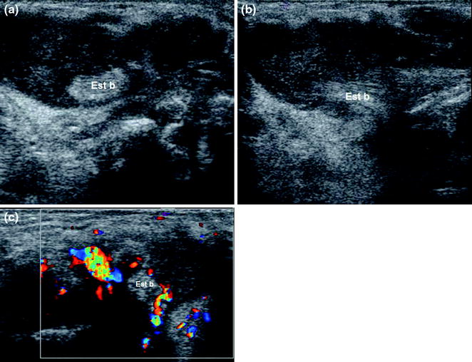
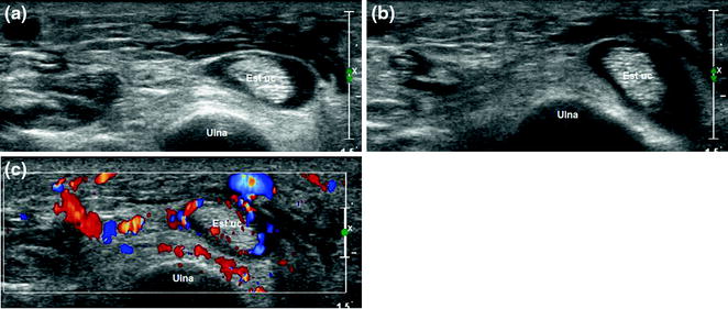
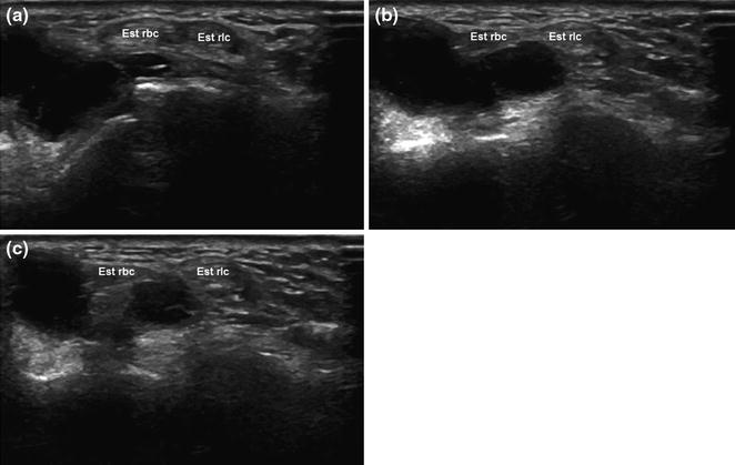
Fig. 3.4
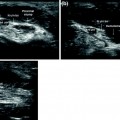
Purulent tenosynovitis involving the first compartment. Sonography reveals corpuscular fluid within the sheath surrounding the abductor longus and extensor brevis of the thumb (Est b) (a, b). Color Doppler imaging shows hypervascularization (c). The patient was generally debilitated, and the tenosynovitis was the result of a minor wound that became infected
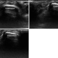
Fig. 3.5
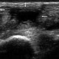
Tenosynovitis of the extensor carpi ulnaris (Est uc). Sonography shows fluid within the tendon sheath, edema (a, b) and hypervascularization (c) of the soft tissues at the site of a recent dog bite
Fig. 3.6


Related posts:
Stay updated, free articles. Join our Telegram channel
Full access? Get Clinical Tree
