Clinical: Deep, painless, firm or hard, and poorly circumscribed mass that may be adherent to the skin or bone and that grows insidiously and slowly for several months. Tumor mass is elongated along the muscle, or it is made up of large masses disseminated along the limb and joined together. The tumor grows by infiltrative ramifications along the muscle, the aponeuroses, and the tendons. When large, muscular retraction phenomena and limited joint motion occur. Pain, numbness, hypoesthesia, and motor weakness when it infiltrates a large nerve.
Imaging: On X-ray a dense mass interrupts the adjacent intermuscular and soft tissue planes. Adherent to bone (37 %) with saucerized cortex without periosteal reaction. On angiography a thick intratumoral capillary network, with few vessels of large caliber. On CT intensely and diffusely takes up contrast medium, easily distinguished from surrounding muscles, irregular shape of the tumor, many nodules joined together, and faded boundaries and digitations along the fascial and interstitial planes. On MRI infiltrative pattern (63 %) in youngsters and nodular pattern (81 %) in adults; hypo-/isointense to muscle on T1 and intermediate on T2; inhomogeneous (75 %), bright in the center (cellular area) and dark in the periphery (collagen area) on T2; on T2 white zones enhance after gadolinium administration, whereas dark zones remain hypointense; bright mass on T2 are aggressive lesions; natural evolution shows proximal migration; and gradual decrease of enhancing cellular areas and increasing of nonenhancing collagen zones.
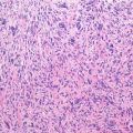
Histopathology: Dense, white, firm lesion that may be of large size (up to 20 cm). Ill-defined margins, digitations, and satellite nodules near the tumoral limits. Firmly adherent to the aponeuroses, tendons, joint capsule, vessels, and nerves. These are compressed but not invaded. Rarely, compression on the bone causes a superficial scalloping of the cortex. A densely collagenized mature fibrous connective tissue with a relatively sparse number of fibroblasts. Mitoses are rare or absent. Variably prominent ectatic blood vessels. Spindle cells in intertwined bundles that are wavy and have no particular order. Infiltration of the muscle and the surrounding tissues. On immunohistochemistry, desmoid tumor is diffusely positive for vimentin and shows nuclear expression of beta-catenin in 85 % of cases. It may be positive for pan-muscle actin and smooth muscle actin.
Related posts:


Stay updated, free articles. Join our Telegram channel

Full access? Get Clinical Tree


