Location: Any soft tissue site can be involved, but most common are the paravertebral region, retroperitoneum, and chest wall, followed by the extremities.
Clinical: Large, destructive mass.
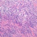
Histopathology: Grossly it is a large, destructive mass, frequently with necrosis and hemorrhage. Histologically, Ewing’s sarcoma shows a sheetlike to vaguely lobular growth pattern, a well-developed capillary vascularization, and a uniform cell population of round blue cells with small amounts of clear to light eosinophilic cytoplasm, regular nuclear contours, finely dispersed chromatin, and small nucleoli. Geographic necrosis and individual apoptotic cells are frequently seen. Pseudorosettes are a consistent clue for diagnosis of PNET. Immunohistochemical and molecular findings are the same as for the bone counterpart.
Course and Staging: 10-year survival rate of 60 % with current treatment regimens.
Treatment: Current multimodal therapy (surgery, chemotherapy, and radiotherapy).
Immunohistochemical Panel
VIM | + |
CD99 | + |
Caveolin-1 | + |
Fli1 | + |
CAM 5.2 | ± |
S100 | ± |
LAC | – |
TdT | – |
Chromosomal Translocations
t (11,22)(q24;q12) | EWSR1-FLI1 | 90–95 % |
t (21;22)(q22;q12) | EWSR1-ERG | 5–10 % |
t (2;22)(q35;q12) | EWSR1-FEV
Related posts:Stay updated, free articles. Join our Telegram channel
Full access? Get Clinical Tree
 Get Clinical Tree app for offline access

|