Location: Deep-seated, proximal extremities and trunk.
Clinical: Enlarging painless soft tissue mass.
Imaging: Although imaging characteristics are nonspecific, most tumors appear lobulated, and highly myxoid tumors have a homogeneous high intensity signal on T2-weighted MRI images. Tumors with necrosis or hemorrhage have a more heterogeneous appearance.
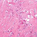
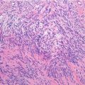
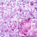
Histopathology: There are cytologically uniform oval and spindle cells embedded in flocculent myxoid ground substance, with fibrous bands dividing individual lobules of tumor. Cells may be arranged as cords, strands, nests, and sheets and may have typically uniform dark-staining nuclei with indistinct nucleoli. In hypervascular matrix-poor regions, tumor cells often display larger, more vesicular nuclei with visible nucleoli; some tumor cells may have distinctly rhabdoid morphology. Tumor is hypovascular and the matrix stains alcian blue positive at pH 4.0 and 1.0 (chondroitin sulfate positive). At the genetic level, the majority of tumors exhibit the t(9;22)(q22;q12) translocation involving EWS and CHN genes.
Course and Staging: Five-year survival rates are high (>80 %); however, late lung metastases occur, and 10- and 15-year disease-free survival rates are considerably lower.
Treatment: Wide surgical excision.
Chromosomal Translocations
t(9;22) (q22;q12) | EWS-NR4A3 (CHN, TEC, NOR1) | 72 % |
t(9;17) (q22;q11)
Stay updated, free articles. Join our Telegram channel
Full access? Get Clinical Tree
 Get Clinical Tree app for offline access

|


