Location: Deep soft tissues of the thigh and buttocks.
Clinical: Progressively enlarging painless mass.
Imaging: X-rays, CT, and MRI reveal a large deep-seated soft tissue mass with variable mineralization. By definition, these lesions do not arise from bone but secondarily involve the periosteum, cortex, or medullary canal.
Histopathology: Highly cellular, mitotically active tumors. There is usually considerable nuclear pleomorphism with necrosis, and lacelike eosinophilic osteoid outlines individual cells or clusters of cells. Mineralized osteoid (bone) is relatively uncommon. Lobules of cellular atypical hyaline cartilage may be present. All histological patterns of osseous osteosarcoma may be seen.
Course and Staging: High-grade tumors, poor prognosis, high rate of metastases.
Treatment: Wide or radical excision with systemic chemotherapy.
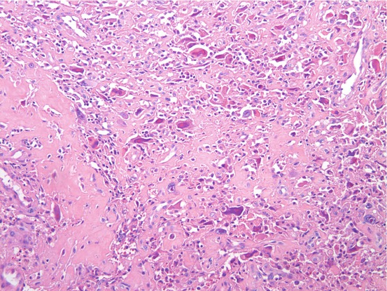
Osteoid-producing cells haphazardly organized, growing in the soft tissue
Selected Bibliography
Goldstein-Jackson SY, Gosheger G, Delling G, Berdel WE, Exner GU, Jundt G, Machatschek JN, Zoubek A, Jürgens H, Bielack SS, Cooperative Osteosarcoma Study Group COSS (2005) Extraskeletal osteosarcoma has a favourable prognosis when treated like conventional osteosarcoma. J Cancer Res Clin Oncol 131(8):520–526PubMedCrossRef
Related posts:


Stay updated, free articles. Join our Telegram channel

Full access? Get Clinical Tree


