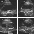
Figure 13.1.1
Thanatophoric dysplasia. A: Sonogram of right lower leg demonstrating bowing of both the tibia (arrow) and the fibula (arrowhead). Both long bones are markedly short for gestational age. B: Image of cranium of another fetus with thanatophoric dysplasia demonstrating a cloverleaf skull, with frontal bossing (arrows) and lateral protrusion in the region of the temporal lobes (arrowheads). C: Longitudinal image of same fetus as (B) showing narrow thorax (arrows) in comparison with larger abdomen (arrowheads). The heart (H) fills most of the thoracic cavity, leaving little room for growth of the lungs.
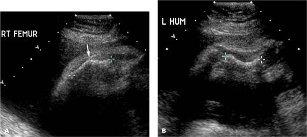
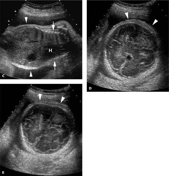
Figure 13.1.2
Osteogenesis imperfecta type II. A: Sonogram of right femur (calipers) with a fracture deformity (arrow), in a fetus with osteogenesis imperfecta type II. B: The left humerus (calipers) is bowed. C: The thorax (arrow) is narrowed, as seen by comparison with the wider, more normal abdomen (arrowheads) (H = heart). (D) and (E) Images of the cranium showing poor ossification, making intracranial contents easy to visualize. The skull (arrowheads) is very soft, such that when gentle compression was applied for image (E), the cranium flattened.
The less severe, nonlethal skeletal dysplasias are often not diagnosable prenatally during the second trimester, because the long bones are not abnormally shortened or deformed at that stage of pregnancy. In the third trimester, measurements of the long bones with these skeletal dysplasias may begin to lag behind the expected size for gestational age, but typically they do not fall more than four standard deviations below the mean. Sometimes, in the third trimester, bowing or fractures (Figures 13.1.5 and 13.1.6) may become apparent, particularly with the nonlethal forms of osteogenesis imperfecta.
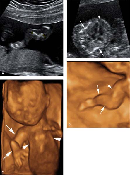
Figure 13.1.3
Osteogenesis imperfecta type II. A: Sonogram of humerus (calipers) showing marked bowing and shortening. B: Axial image of chest showing thorax narrowed by fractured indented ribs (arrows). The heart (arrowhead) fills much of the thoracic cavity. C: 3D image of fetus demonstrating very narrow thorax (arrowheads) and deformity of the right upper extremity (arrows). D: 3D image of right upper extremity showing shortening of the upper arm (arrowhead) and bowing of the forearm (arrows).
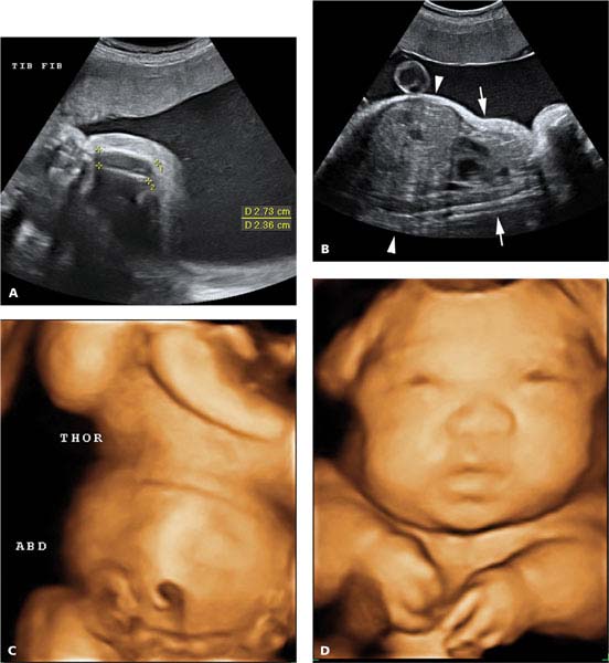
Figure 13.1.4
Achondrogenesis. A: Longitudinal image of lower leg showing shortened and poorly mineralized tibia and fibula (calipers). B: Sagittal image of fetus showing very narrow thorax (arrows) compared to the abdomen (arrowheads). C: 3D image of trunk of fetus showing very small thorax (THOR) compared to the abdomen (ABD). D: 3D image of face and upper trunk demonstrating the very short upper extremities.
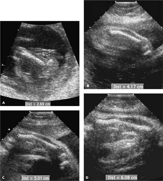
Figure 13.1.5
Osteogenesis imperfect type I with development of bowing. Femur measurements (calipers) at (A) 17 weeks, (B) 25 weeks, (C) 29 weeks, and (D) 33 weeks showing straight femur at 17 weeks, mild bowing at 25 weeks, and moderate bowing at 29 and 33 weeks. At 17 weeks the femur length was above the mean for gestational age. From 25 weeks onward, the femur length fell below the mean for gestational age, but never more than two standard deviations below.

Figure 13.1.6
Osteogenesis imperfect type I with bowing of long bones. Images of (A) right femur (arrowheads) and (B) right tibia (calipers) showing bowing of both bones. C: 3D sonogram of forearm and hand with skeletal settings demonstrating angulation of the distal ulna and radius (arrowheads).
13.2. Skeletal Dysostoses
Description and Clinical Features
Defective ossification of one bone or a group of bones is called a skeletal dysostosis. Some skeletal dysostoses have a recognizable pattern of deformities and are part of a known syndrome, such as Nager acrofacial dysostosis, Poland syndrome, and proximal focal femoral deficiency. Nonskeletal anomalies are common in many of these syndromes. Other dysostoses are isolated bony deformities with no known cause and no other fetal anomalies. The prognosis for skeletal dysostoses is related to the degree of severity of other anomalies and the extent of osseous deformities. Deformities of the head, spine, and thorax carry a worse prognosis than deformities isolated to the extremities.
Sonography
The sonographic appearance of a skeletal dysostosis depends on the bony structures involved. For example, with Nager acrofacial dysostosis, the upper extremities are markedly shortened, with absence of one or more of the long bones. The hands are present but incompletely formed. A hypoplastic mandible and external ear deformities are also present (Figure 13.2.1). Proximal focal femoral deficiency is characterized by the absence of the proximal femur. The finding is unilateral in 90% of cases. The diagnosis is made when there is extreme shortening of one femur with a normal contralateral femur. Other skeletal anomalies may be present, most commonly in the affected lower extremity (Figure 13.2.2).
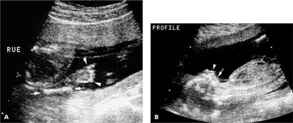
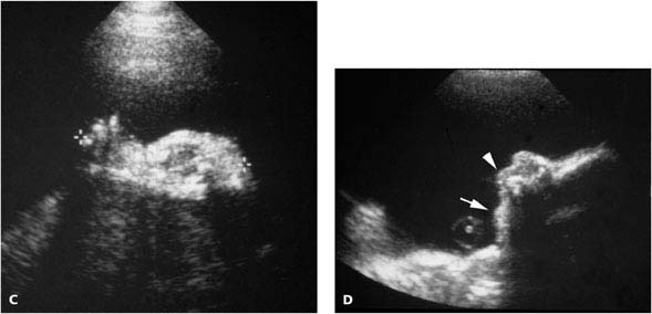
Figure 13.2.1
Nager acrofacial dysostosis. A: Right upper extremity (RUE) is abnormally formed, with absence of the bones of the forearm such that the hand (arrowheads) is contiguous with a short humerus (arrows). B: The mandible (arrow) is markedly hypoplastic beneath the normally formed maxilla (arrowhead), visible on a profile of the face. C: Image in another fetus with Nager acrofacial dysostosis demonstrating an extremely short upper extremity (calipers) due to absence of the bones of the forearm. D: Sonogram of facial profile in same fetus as (C) showing markedly hypoplastic mandible (arrow) with normally formed maxilla (arrowhead).
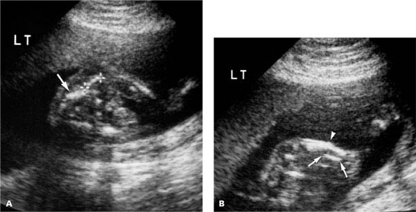
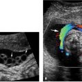
Figure 13.2.2
Proximal focal femoral deficiency with abnormal lower-extremity long bones. A: The femur (calipers), seen adjacent to the iliac crest (arrow), is very short. The contralateral femur was normal. B: The fibula (arrows) in the leg with the short femur is hypoplastic and the tibia is bent midshaft (arrowhead).
Related posts:


Stay updated, free articles. Join our Telegram channel

Full access? Get Clinical Tree


