Figure 18.1.1
Oligohydramnios in the first trimester. There is little fluid around this 7-week embryo (calipers). The fetus died within 3 weeks of this sonogram.
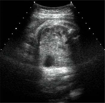
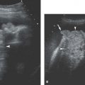
Figure 18.1.2
Oligohydramnios in the third trimester diagnosed by subjective assessment. The amniotic fluid volume appears subjectively low in this 32 weeks gestation sonogram.
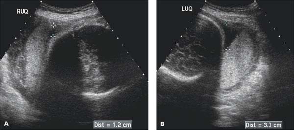
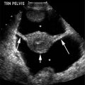
Figure 18.1.3
Oligohydramnios in the third trimester diagnosed by amniotic fluid index (AFI). Deepest pocket measurements in (A) right upper quadrant (RUQ), (B) left upper quadrant (LUQ), (C) right lower quadrant (RLQ), and (D) left lower quadrant (LLQ). AFI is calculated as the sum of these measurements and yields an amniotic fluid index of 4.2 (1.2 + 3.0 + 0 + 0), indicating oligohydramnios because it is <5 cm.
18.2. Polyhydramnios
Description and Clinical Features
Excessive amniotic fluid volume, or polyhydramnios, can occur with a number of maternal and fetal abnormalities. Diabetic mothers have a higher frequency of polyhydramnios than do nondiabetic mothers. Fetal anomalies that impair swallowing or decrease gastrointestinal tract absorption of amniotic fluid also lead to polyhydramnios. These anomalies include esophageal, duodenal, and proximal small bowel obstruction; severe brain anomalies such as anencephaly; facial clefts and tumors; and neck and intrathoracic masses. Hydropic fetuses are often surrounded by excessive amniotic fluid. Polyhydramnios may also be idiopathic in that no maternal or fetal cause is identified.
Polyhydramnios, regardless of its cause, may lead to various maternal symptoms. The mother may experience pain or premature uterine contractions as a result of excessive stretching of the uterus. Pressure by the enlarged uterus on adjacent structures can cause maternal hydronephrosis, lower extremity edema, and respiratory problems.
Sonography
The sonographic diagnosis of polyhydramnios in the second and third trimesters can be established by subjective assessment of amniotic fluid volume, deepest pocket measurement, or AFI (Figures 18.2.1 and 18.2.2). A deepest pocket measurement >8–10 cm or an AFI >18–20 cm is generally considered diagnostic of polyhydramnios.
When polyhydramnios is diagnosed, a careful fetal anatomic survey should be undertaken to search for a cause. In particular, the fetal head, face, neck, thorax, and gastrointestinal tract should be evaluated.
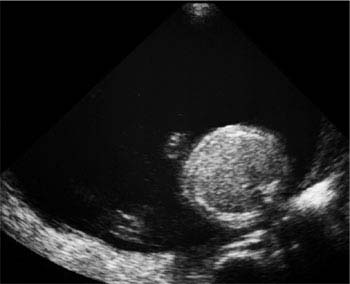
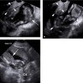
Figure 18.2.1
Polyhydramnios: subjective assessment. The volume of amniotic fluid appears markedly increased. No stomach is seen in the fetal abdomen, because the fetus has esophageal atresia.
Related posts:


Stay updated, free articles. Join our Telegram channel

Full access? Get Clinical Tree


