 Figure 6.1.1
Unilateral cleft lip on grayscale ultrasound. A: Coronal image of lower face demonstrating large unilateral defect (long arrow) in the upper lip (short arrows) extending into the ipsilateral nostril. The lower lip (arrowheads) is seen inferior to the cleft. B: Coronal image of the same fetus slightly more anterior demonstrating large fluid-filled cleft (*) in the upper lip (arrows) above the lower lip (arrowheads). C: Axial image of the upper lip showing defect (arrow) on the left (R, right; L, left).
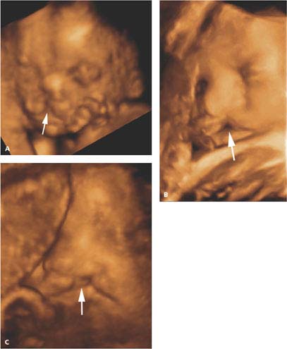
Figure 6.1.2
Three-dimensional (3D) ultrasound of unilateral cleft lip. A, B, and C: 3D images of fetal face demonstrating defect in the upper lip (arrow).
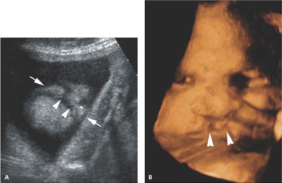
 Figure 6.1.3
Bilateral cleft lip. A: Image of bilateral clefts (arrowheads) in the upper lip (arrows). B: Three-dimensional image in the same fetus as in (A) showing bilateral clefts (arrowheads).
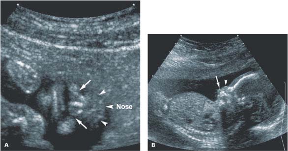
 Figure 6.1.4
Bilateral cleft lip and palate with maxillary prominence. A: Coronal image of lower face demonstrating bilateral clefts in the upper lip (arrows) on either side of bony protuberance, representing the maxillary prominence, inferior to the nose (Nose, arrowheads). B: Sagittal image of face demonstrating maxillary protuberance (arrow), extending farther from the face than the nose (arrowhead).
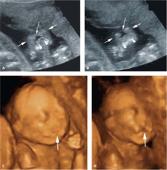
Figure 6.1.5
Midline facial cleft. A and B: Coronal images of mouth demonstrating a central defect (long arrow) in the upper lip (arrows). The lower lip (arrowheads) is inferior to the cleft. C and D: Three-dimensional images of midline cleft lip (arrow) extending into the nose.
6.2. Macroglossia
Description and Clinical Features
Macroglossia is an abnormally large tongue. It can occur in fetuses of diabetic mothers, as well as in fetuses with various syndromes, including Beckwith-Wiedemann (a syndrome characterized by organomegaly and omphalocele), trisomy 21, and congenital hypothyroidism. The enlarged tongue protrudes from the mouth and may interfere with swallowing, thus causing polyhydramnios.
Sonography
When macroglossia is present, the tongue persistently protrudes from the fetal mouth (Figures 6.2.1 and 6.2.2). When the tongue is seen protruding from the mouth, careful examination of the fetal mouth is warranted to determine whether the tongue itself is enlarged or the tongue is being displaced by an oral mass (see Figure 6.7.3). Once the diagnosis of macroglossia is made, the fetus should be assessed for other anomalies, such as omphalocele, which is associated with Beckwith-Wiedemann syndrome.
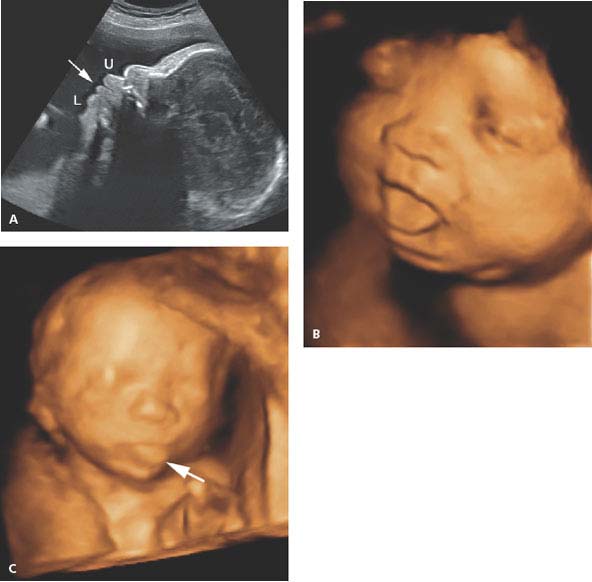
Figure 6.2.1
Macroglossia. A: Sagittal image of face demonstrating large tongue (arrow) protruding anteriorly between upper (U) and lower (L) lips. B: Three-dimensional (3D) image of the same fetus showing protruding tongue. C: 3D image in a fetus with trisomy 21 and enlarged tongue (arrow) seen protruding from the mouth.
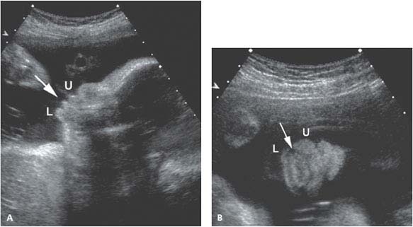
 Figure 6.2.2
Macroglossia in a fetus with Beckwith-Wiedemann syndrome. Sagittal (A) and coronal images (B) of face in a fetus with Beckwith-Wiedeman syndrome demonstrating large tongue (arrow) protruding anteriorly between upper (U) and lower (L) lips.
6.3. Micrognathia
Description and Clinical Features
Micrognathia is defined as a small or hypoplastic mandible. This anomaly is associated with multiple syndromes and chromosomal abnormalities. In particular, it is a common finding in fetuses with trisomy 18 and trisomy 13. Micrognathia is a component of various skeletal dysplasias and dysostoses, such as Treacher Collins syndrome and Nager acrofacial dysostosis.
The presence of a small mandible may interfere with fetal swallowing. As a result, micrognathia may lead to polyhydramnios.
Sonography
The small mandible, characteristic of micrognathia, is best identified sonographically on a sagittal midline view of the fetal face demonstrating a small chin or with 3D imaging (Figure 6.3.1). Because of the association of micrognathia with aneuploidy, careful evaluation of the fetus is warranted once a small mandible is identified to look for sonographic findings associated with abnormal chromosomes.

Figure 6.3.1
Micrognathia. A: Sagittal profile of face demonstrating very small mandible and chin (arrow). Sagittal image (B) and three-dimensional image (C) of another fetus with small mandible and chin (arrow).
Related posts:


Stay updated, free articles. Join our Telegram channel

Full access? Get Clinical Tree


