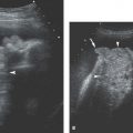
Figure 25.1.1
Normal ovary in a woman of menstrual age. The ovary (arrowheads) appears as a structure of moderate echogenicity, containing several small functional cysts (*’s).
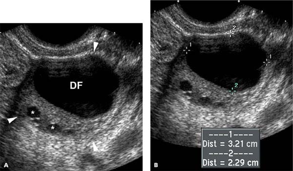
Figure 25.1.2
Normal ovary with a dominant follicle in a woman of menstrual age. A: The ovary (arrowheads) contains several small functional cysts (*’s) and one much larger cyst representing the dominant follicle (DF). B: There are cursors measuring the dominant follicle.
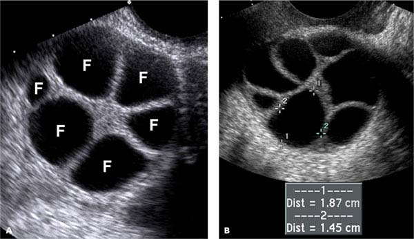
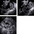
Figure 25.1.3
Ovary in a woman undergoing treatment for infertility. A: In this woman taking medication to stimulate development of ovarian follicles, ultrasound demonstrates multiple follicles (F) throughout the ovary. These occupy a relatively larger portion of the ovary than they do in a normal, nonstimulated ovary. B: Follicle measured (calipers) in another women taking medication to stimulate follicular development.
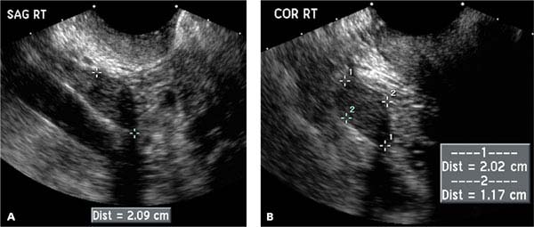
Figure 25.1.4
Normal ovary in a postmenopausal woman. A: Sagittal and (B) coronal transvaginal images demonstrate the right ovary (calipers) in a postmenopausal woman. The ovary is small and homogeneous, without the physiologic cysts seen in the typical premenopausal ovary.
Related posts:


Stay updated, free articles. Join our Telegram channel

Full access? Get Clinical Tree


