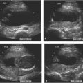
Figure 19.1.1
Cross section of two-vessel umbilical cord. A: Image of cord surrounded by amniotic fluid demonstrating two vessels. The larger one is the umbilical vein (arrow) and the smaller is the single umbilical artery (arrowhead). B: Color Doppler demonstrating red in the umbilical vein (arrow) and blue in the umbilical artery (arrowhead), demonstrating that flow is in opposite directions in the two vessels.
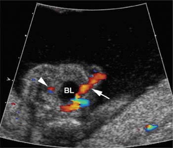
Figure 19.1.2
Single umbilical artery in pelvis. Color Doppler transverse image of pelvis demonstrating a single umbilical artery (arrow) adjacent to one side of the bladder (BL). On the contralateral side, no umbilical artery is seen arising from the iliac artery (arrowhead).
19.2. Abnormal Placental Cord Insertions and Vasa Previa
Description and Clinical Features
Marginal and velamentous insertions of the umbilical cord into the placenta are abnormal placental cord insertions associated with increased risks to the fetus. A marginal cord insertion is one that enters the placenta within 1 cm of the placental edge. A velamentous cord insertion is present when the umbilical cord terminates at the uterine wall distant from the placenta, with the umbilical vessels traveling beneath the membranes before inserting into the edge of the placenta. Predisposing factors for these abnormal insertions include multiple gestations, a low-lying placenta, placenta previa, uterine anomalies, and uterine scarring. Risks include cord rupture and thrombosis.
Marginal cord insertions are thought to develop during the course of gestation as the placenta evolves. In particular, if placental tissue on one side of the cord atrophies while placental tissue on the other side of the cord grows, the position of the cord in the placenta will be closer to one edge of the placenta than the other.
Velamentous cord insertions are thought to develop when there is atrophy of placental tissue on one side of the cord as well as placental tissue beneath the cord insertion site, leaving the umbilical vessels exposed beneath the membranes as they travel to the remaining placental mass. If the vessels of a velamentous umbilical cord travel across the cervix beneath the membranes, the configuration is termed vasa previa.
Vasa previa may also result when the placenta has a succenturiate or accessory lobe. Succenturiate lobes are connected to the rest of the placenta by fetal vessels. When these connecting fetal vessels course across the cervix, a vasa previa is present.
Vasa previa carries a high risk of perinatal mortality, because the fetal umbilical vessels coursing over the cervix are prone to bleeding, especially if the vasa previa is not diagnosed prenatally and vaginal delivery is attempted.
Sonography
Both color Doppler and gray-scale ultrasound are useful for detecting and characterizing abnormal umbilical cord insertions. With a marginal cord insertion, the umbilical cord insertion into the placenta is seen near the edge of the placenta (Figure 19.2.1). With velamentous cord insertion, the intra-amniotic umbilical cord is seen terminating at the uterine wall away from the placenta, with the umbilical vessels traveling beneath the membranes to the placenta (Figure 19.2.2).
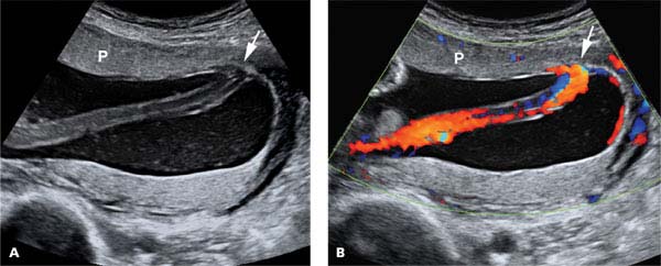
Figure 19.2.1
Marginal umbilical cord insertion. A: Sonogram showing umbilical cord inserting into the placenta (P) at its edge (arrow). B: Color Doppler demonstrating placental cord insertion (arrow) close to the edge of the placenta (P).
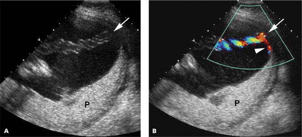
Figure 19.2.2
Velamentous cord insertion in one of twins. A: Sonogram demonstrating umbilical cord insertion (arrow) into uterine wall away from the placenta (P) in one of a twin gestation. B: Color Doppler image showing velamentous cord insertion (arrow) with the umbilical vessels (arrowhead) traveling beneath the membranes to the placenta (P).
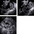
Vasa previa is diagnosed when the vessels of a velamentous cord (Figure 19.2.3), or vessels connecting a succenturiate lobe to the main placenta, are seen crossing the cervix. Transvaginal scanning may be necessary to visualize these abnormal vessels in cases where the fetal head obscures the cervix (Figure 19.2.4). Care must be taken to distinguish a vasa previa, in which the cord is held in place over the cervix by the overlying membranes, from a cord that is free floating but held over the cervix by the fetal head. When the distinction between these is uncertain, the correct diagnosis can be made by trying to lift the fetal head manually or by seeing if the cord remains over the cervix on a follow-up scan.
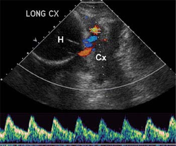
Figure 19.2.3
Vasa previa on transabdominal scan. Sagittal image of lower uterine segment with color and spectral Doppler showing blood flow in vessels between the fetal head (H) and cervix (Cx). The spectral Doppler demonstrates an umbilical artery waveform, proving these are umbilical vessels of a vasa previa.
Related posts:


Stay updated, free articles. Join our Telegram channel

Full access? Get Clinical Tree


