This is a standard view for looking at the shoulder joint. The bones, joints, lung and soft tissues are visible. The surgical neck of the humerus is a common site of proximal humeral fracture. The GHJ is perfectly aligned. GHJ glenohumeral joint, ACJ acromioclavicular joint, GT greater tubercle, LT lesser tubercle (of humerus)
20.2 Normal shoulder: Y-view
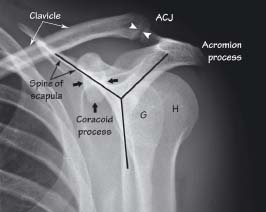
On this view the scapula forms a Y shape comprising the body, spine and acromion process. The humerus (H) overlies the glenoid (G) and lies behind the coracoid process. ACJ acromioclavicular joint
20.3 Normal shoulder: axial view
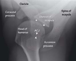
This view is taken with the patient’s arm abducted, but is often not tolerated by the patient if there is restriction of movement due to pain. The view gives a clear indication of glenohumeral joint (*) alignment. The head of the humerus is seen to lie behind the coracoid process
20.4 Normal elbow – 13-year-old boy: lateral and AP views
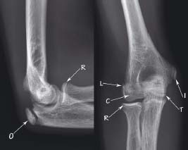
The ossification centres of the elbow are demonstrated. These form the mnemonic ‘CRITOL’ according to the order of their appearance. C capitulum, R radial head, I internal (medial) epicondyle, T trochlea, O olecranon, L lateral epicondyle
20.5 Normal hand: oblique and AP view
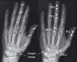
M metacarpal bone, PP proximal phalanx, MP middle phalanx, DP distal phalanx, CMCJ carpometacarpal joint, IPJ interphalangeal joint (thumb only), MCPJ metacarpophalangeal joint, PIPJ proximal interphalangeal joint, DIPJ distal interphalangeal joint
20.6 Normal wrist/carpal bones: AP with ulnar angulation
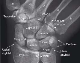
In this view the wrist is stressed towards the ulnar side in order to ‘open’ any fracture present in the scaphoid. CMCJ carpometacarpal joint, RCJ radiocarpal joint, DRUJ distal radio-ulnar joint, TFCC position of triangulofibrocartilage complex, M metacarpal bone
Upper limb anatomy seen on XR
Plain X-ray imaging demonstrates the normal bony anatomy of the limbs very well, however, normal soft tissue structures are less well seen. The important bony landmarks are centred around the joints. The anatomical position is used for description (standing with palms forwards). The five principal densities seen on plain imaging (see Chapter 1) should be used as reference to identify anatomical and pathological structures.
Shoulder
Related posts:


Stay updated, free articles. Join our Telegram channel

Full access? Get Clinical Tree


