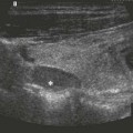
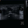
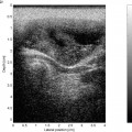
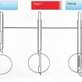
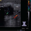
10 Facial Soft Tissues The hypoechoic structure of the masseter muscle with its pinnate reflection pattern is a primary landmark in the transverse view of the parotid region. The parotid duct (Stensen duct) lies superficially to the muscle but is usually only identifiable as a hypoechoic/anechoic area when it is obstructed (Fig. 10.1). The duct runs anteriorly and medially, traverses the buccal fat pad, and opens into the vestibule of the mouth, opposite the second upper molar, after passing through the (hypoechoic) band of the buccinator muscle (Fig. 10.2). The orbicularis oris muscle and the soft tissues of the lip can be seen anterior to the oral cavity region. These tissues are divided into three layers: a central hypoechoic structure with a more echogenic band in front and behind, corresponding to the anatomical structure of the lips with two layers of subcutaneous tissue and the orbicularis oris muscle (Fig. 10.3). When examining facial structures such as the cartilaginous structures of the auricle, mastoid, or face, ultrasound gel should be applied in sufficient amount to ensure good acoustic conditions. Anterior to the parotid gland and lying on the ascending ramus of the mandible (echogenic reflection with distal acoustic shadowing), is the masseter muscle with its characteristic pinnate reflection pattern. It can be displayed in the longitudinal and transverse planes, as well as in relaxation and in maximum contraction. Masseter hypertrophy: Apart from a condition affecting the parotid gland, unilateral swelling of the cheek may be due to hypertrophy of the masseter muscle caused by an occlusal parafunction (Fig. 10.4). The temporomandibular joint is also accessible to ultrasound. The dynamics of the joint can be examined to determine, among other things, any dislocation or disorder of movement ( A furuncle is an infection of a hair follicle with surrounding tissue reaction; it may occur anywhere on skin with hair. With the typical clinical external picture, ultrasound imaging shows a hypoechoic, star-shaped, loosely structured lesion in the skin (Fig. 10.5). The extent of the inflammatory reaction in the deeper tissues can easily be assessed. Anechoic areas with distal acoustic enhancement indicate an abscess (Fig. 10.6). Color-coded duplex sonography (CCDS) characterizes the inflammatory hyperperfusion (Figs. 10.7, 10.8). Pearls and Pitfalls Furuncles above the oral fissure require assessment with Doppler sonography or color duplex imaging of the angular vein. Any signs of venous thrombosis (Fig. 10.9) mean that there is a danger of intracranial spread. Retroauricular swelling with a prominent ear on inspection correlates with the ultrasound appearance of a subcutaneous hypoechoic thickening (Fig. 10.10). If the ongoing inflamamation breaches the mastoidal osseous cortex the abscess can easily be recognized as a gap in the continuity of the cortical bone (Fig. 10.11). A rapid diagnosis can be made with ultrasonography, especially in children. Ultrasound examination may be worthwhile in patients, especially children, after lateral skull base surgery such as cochlear implantation and/or for local wound monitoring (Fig. 10.12; Preauricular sinuses and auditory meatal fistulas are ectodermal tissue inclusions that can sometimes be assessed on ultrasound imaging with respect to their depth and branching (Figs. 10.13, 10.14). When planning the extent of surgery, particular attention must be paid to the possibility of a rare intraparotid extension. Pearls and Pitfalls If the fistula can be probed, it may be worth trying a careful instillation of hydrogen peroxide, as a contrast enhancer, to delineate the lesion more clearly in the deeper tissues. Epidermoid cysts or trichilemmal cysts, arising as a result of an obstruction of sebaceous ducts, are ubiquitous on the hair-covered scalp. The adipocytes, fat crystals, and epidermal cells that comprise these lesions generate an inhomogeneous, hypoechoic picture. In the noninflammatory stage, the epidermoid cyst is clearly demarcated from its surroundings and there is often the suggestion of a hypoechoic duct (Figs. 10.15, 10.16, 10.17). As well as an increase in volume, inflammation causes a loss of marginal definition and hyperperfusion. Anechoic areas with distal acoustic enhancement indicate abscess formation (Fig. 10.18). Depending on the echotexture, ultrasonography can indirectly determine the consistency of a new lesion. As is the case with cavities filled with viscous mucus, mucoceles are firm, elastic, well demarcated, and mobile on sonographic palpation, and display absolutely no intrinsic vascularity (Fig. 10.19). Posttraumatic hematomas of the face can be seen as hypoechoic, usually subcutaneous, irregularly defined space-occupying lesions without any signs of intrinsic perfusion (Fig. 10.20). Associated with an increasing organization of the hematoma, vascularization can be seen on CCDS over the course of time (Figs. 10.21, 10.22). Solid tumors with visible intrinsic perfusion tend to be hypoechoic or inhomogeneously echoic. To a limited extent, the consistency of a lesion or swelling can also be estimated on the basis of its echogenicity and the findings on sonographic palpation (Figs. 10.23, 10.24, 10.25; At first glance, the different types of vascular malformations have a similar abnormal ultrasound features in B-mode scans, which makes differentiation more difficult. There is a compressible, loosely structured, honeycomb echo pattern that is partly hypoechoic, partly echogenic (Figs. 10.26, 10.27). Ultrasonography allows the assessment of the extent and depth of these lesions in the relevant structure and adjacent soft tissues. The use of color Doppler ultrasound makes it possible to differentiate vascular malformations from other masses (Figs. 10.28, 10.29). Hemangiomas have a heterogeneous echotexture that is hypoechoic with sinusoidal compartments. The perfused areas of these angiomas are, however, visible on CCDS and measurements can be taken to assess venous and arterial elements (Figs. 10.30, 10.31, 10.32). The afferent vessels are branches from the carotid artery territory. Echogenic internal echoes indicate intrinsic venous calcification (phleboliths). Compression with the probe induces an increase in the velocity of the Doppler flow curve. Lymphangiomas/hygromas, on the other hand, do not produce any color flow or Doppler signals and are easily compressible. Their characteristic feature is a loculated appearance that makes it possible to classify them as macrocystic or microcystic lesions (Figs. 10.33, 10.34). With respect to location, most lymphangiomas are found at submandibular, supraclavicular, or parotid sites (see also Chapter 6, p. 75, and Chapter 10, p. 152). The role of ultrasound-guided fine-needle aspiration biopsies and percutaneous and interstitial laser therapy must be mentioned. It may also be used when injecting sclerotic agents under direct visual.
Anatomy
 Video 10.1).
Inflammatory Changes
Furuncles and Abscesses
Mastoiditis
 Video 10.2).
Preauricular Sinus
Benign Tumors
 Videos 10.3, 10.4).
Malignant Tumors
Related posts:
Stay updated, free articles. Join our Telegram channel

Full access? Get Clinical Tree


