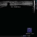
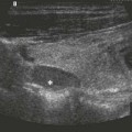
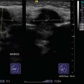
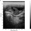
12 Larynx and Hypopharynx The larynx and hypopharynx are not always optimally accessible to ultrasound examination because of their anatomical position, with surrounding air-filled spaces and the irregular ossification of the thyroid cartilage found in adults. As a rule, the retropharyngeal space cannot be assessed completely (even when the probe is tilted) because of its location behind the pharynx. In children and adolescents, however, the examination usually gives a complete overview of the structures in this area. The area should be examined with the patient’s head reclined and with sufficient gel applied to even out the irregular contours of the laryngeal prominence. Starting with the acoustic shadowing of the hyoid, the preepiglottic space can be assessed in cross-section (Fig. 12.1). In the transverse plane, the epiglottis is visible as a horizontal arching hypoechoic band. Moving the probe parallel to the midline, the spindle-shaped/oval, hypoechoic, and finely pinnate prelaryngeal muscles, such as the thyrohyoid, sternohyoid, and sternothyroid, can be seen in cross-section. Below these, the tentlike alae of the thyroid cartilage can be seen on both the right and the left (Figs. 12.2, 12.3; In the endolarynx, it is not always possible to identify the vestibular folds as individual structures with any certainty, as the paraglottic fat and adjacent sinus of Morgagni, with its dense air echoes, may be superimposed (Fig. 12.4; On quiet respiration, the vocal cords can be distinguished caudally as echogenic lines converging anteriorly; they oscillate when the patient phonates ( In their triangular configuration, the arytenoid cartilages can be seen as hypoechoic structures forming the posterior limit of the vocal cords. If conditions are favorable, the epithelial borders of the piriform sinus and the postcricoid region can be identified posterior to the arytenoid cartilage (Fig. 12.5; In adults, it may sometimes be better to examine the larynx unilaterally with the linear array transducer in a paramedian transverse position. Pearls and Pitfalls Asking the patient to say quietly a long “heee” establishes the correct positioning of the vocal cord plane for comparison of oscillation on the two sides. The probe has to be readjusted because of the elevation of the larynx with phonation. Located inferiorly and interrupted by the cricothyroid ligament, the cricoid cartilage, which lies below the thyroid cartilage, and the first tracheal ring can be differentiated (Figs. 12.6, 12.7). Only the superficial part of the trachea can be seen on ultrasound imaging: further down, air prevents the structures within the trachea from being demonstrated clearly. The surface of the trachea can easily be identified by its characteristic “rope ladder” appearance. The cervical esophagus can be identified from its ring-shaped structure and its position, which, anatomically, is immediately to the left of the thyroid gland (Figs. 12.8, 12.9; Pearls and Pitfalls Asking the patient to swallow helps to positively identify the esophagus. The mixture of saliva and air can be seen briefly on its way to the stomach as a pathway of mobile hyperechoic “sparkling” echoes the acoustic shadows caused by scattering. The postion of the esophagus usually moves from the left to the right when the patient’s head is turned to the right. Parapharyngeal and retropharyngeal abscesses are presented in detail in Chapters 6 and 8 (pp. 68–75 and 98). Although laryngoscopy is usually sufficient to diagnose vocal cord polyps and cysts, anterior lesions can be clearly demonstrated as smooth-walled hypoechoic masses. Vocal cord paresis obviously remains a laryngoscopic diagnosis, even if it can be seen on ultrasound scanning as asymmetrical movements when comparing the two sides. Laryngoceles are spaces filled with liquid or mucoid secretions and have to be distinguished from cysts in the neck (Fig. 12.10). The detection of a connection between the endolarynx and upper edge of the thyroid cartilage makes the diagnosis easier. Hypoechoic appearance and lack of any intrinsic perfusion undermine the suspicion of a laryngocele, as does the possible enlargement of the lesion with a Valsalva maneuver. In addition to their detection on laryngoscopy, vascular malformations can also be seen on ultrasonography as moderately to strongly perfused space-occupying lesions with inhomogeneous echogenicity and irregular margins (Figs. 12.11, 12.12). Chondromas are rare space-occupying lesions of the thyroid or cricoid cartilages; they can be seen on ultrasound examination as solid thickenings of the cartilage concerned. In the thyroid cartilage, they more often appear as hypoechoic circumscribed swellings with clearly defined margins (Fig. 12.13). A Zenker diverticulum or pharyngeal pouch can be recognized as a hyperechoic space-occupying lesion with distal acoustic shadowing in the typical site to the left of the thyroid (Figs. 12.14
Anatomy
 Video 12.1). If the cartilage is ossified, its appearance is hyperechoic with distal acoustic shadowing, which may compromise the view of the intrinsic structures of the larynx.
 Video 12.2).
 Videos 12.3, 12.4).
 Video 12.5). The borders of the lateral hypopharyngeal walls extend outward like lips lateral to the caudal thyroid cartilage as a hypoechoic/echogenic double contour.
 Video 12.6). Deeper, the esophagus lies on the spine. Osteophytes that narrow the esophageal lumen and cause dysphagia can also be demonstrated on ultrasonography.
Inflammatory Changes
Benign Tumors
![]()
Stay updated, free articles. Join our Telegram channel

Full access? Get Clinical Tree


