Female Genital Tract


Female Genital Tract


Sharon W. Gould

Sabah Servaes

Edward Y. Lee

José Ernesto Lipsich

Mohamed Issa Tawil

Sara O. Vargas

Monica Epelman


INTRODUCTION

Evaluation of the female genital tract in the pediatric population presents a unique set of diagnostic challenges due to the developmental changes that occur during childhood and adolescence as well as to the cyclic changes that occur with the onset of menses. This chapter reviews up-to-date imaging techniques for evaluating the female genital tract in the pediatric population. In addition, the normal appearance of the pediatric female reproductive organs as well as the imaging findings of important congenital and acquired pediatric female genital tract disorders are discussed.


IMAGING TECHNIQUES


Radiography

Although ultrasound is usually the initial imaging modality of choice for evaluating the pediatric female genital tract, radiography may be performed first in the setting of acute pelvic or right lower quadrant abdominal pain.1 A supine anteroposterior (AP) radiograph from the diaphragm to the upper femurs is most often acquired for evaluating abdominopelvic pain, and it may be complemented with an upright or decubitus view. Radiography may show mass effect on gas-filled bowel loops from pelvic masses and mass-like abnormalities, calcification, or even a tooth in the setting of an ovarian teratoma. Such findings can help generate a differential diagnosis and guide subsequent imaging studies for further evaluation.


Ultrasound

The primary imaging modality for assessment of the pediatric female genital tract is ultrasound due to its multiplanar imaging capability, high soft tissue contrast resolution, and lack of ionizing radiation.1,2 In addition, sedation is typically not required for ultrasound imaging, even in very young children. The ultrasound examination of the female genital tract is usually performed through a fluid-filled bladder using transabdominal technique.1,3,4 To achieve optimal bladder filling, cooperative children can be asked to drink water prior to imaging while refraining from voiding. If the child is uncooperative or the need for evaluation is urgent, the bladder can be catheterized and filled retrograde with sterile saline. Although transvaginal ultrasound technique excellently depicts the female genital tract, it is generally not appropriate in younger children or adolescents who are not yet sexually active.1 Transperineal scanning can assess disorders of the vagina, urethra, and vulva.

A complete ultrasound examination of the pediatric female genital tract includes assessment of the upper portion of the vagina and uterus (including cervix). The width of the endometrial stripe, including both layers, should be recorded; a slightly deeper hypoechoic myometrial layer, rarely seen in pediatric patients, should not be included in the measurement1,3 (Fig. 19.1). Assessment of the adnexae includes measurements of the ovaries in three planes. Ovarian volumes can be calculated and compared to normative values. Note should be made of the presence, appearance, and amount of fluid in the cul-de-sac (between the uterus and rectum), as well as within each lower quadrant of the abdomen. In the setting of acute pelvic pain and possible ovarian torsion, color and spectral Doppler evaluation of the ovaries should be performed with documentation of arterial and venous spectral waveforms.


FIGURE 19.1 Longitudinal ultrasound images through the midline pelvis show the normal appearances of the uterus in various developmental stages. A: Neonate with a relatively large uterus with a well-delineated endometrial stripe under the influence of maternal hormones. Notice the relative prominence of the cervix (arrowheads) with respect to the body and fundus (arrow). B: Prepubertal, tubular appearance of the uterus (arrows) in a 7-year-old girl. No endometrial stripe is seen. The uterus is homogeneously hypoechoic. C: Inverted pear-shaped appearance of the postpubertal uterus. Notice the typical appearance of the endometrial stripe in the periovulatory phase. The thin hypoechoic layer just outside of the calipers represents the inner myometrial layer and should not be included in the endometrial measurement. A small amount of free fluid (asterisk) is present in the cul-de-sac.


Computed Tomography

Computed tomography (CT) generally provides less soft tissue differentiation than ultrasound or magnetic resonance imaging (MRI) and, therefore, is generally less helpful for evaluating the pediatric female genital tract. Radiation exposure associated with CT is also a limitation in children. In addition, CT lacks the “real-time” capability of ultrasound. However, depending on a child’s clinical presentation and institutional protocol, CT may be the first imaging study acquired, especially in the setting of acute right lower quadrant abdominal pain. CT is also used to further characterize and stage masses arising from the pelvis, particularly when malignancy is suspected. In the CT evaluation of pelvic structures, intravenous and oral contrast materials help with lesion characterization as well as distinguishing true pelvic abnormalities from adjacent bowel loops. Single-phase CT imaging is usually sufficient for evaluation of female genital tract anomalies and abnormalities.


Magnetic Resonance Imaging

MRI is an excellent imaging modality for assessing the pediatric female genitourinary tract, particularly for delineation of congenital anomalies5 and characterization of pelvic masses.6,7 MRI’s inherent multiplanar imaging capability and high soft tissue contrast resolution are especially helpful for evaluating the pelvic organs; however, the need for sedation or general anesthesia in younger pediatric patients because of long imaging times limits MRI to a secondary role after pelvic ultrasound.1 Imaging of the pediatric female pelvis should be performed in three planes or with an isotropic 3D imaging protocol that allows diagnostic multiplanar reformatted images. An advantage of 3D imaging is that reformatted images can be created in any oblique plane based on the patient’s anatomy without additional imaging time.

MRI is the modality of choice for detailed assessment of uterine anatomy because images can be acquired in both the uterine long- and shortaxes.5 Hence, MRI is ideal for characterizing suspected Mullerian anomalies, although it may need to be performed in the postpubertal period for optimal diagnosis when the uterus is more developed. T2-weighted pulse sequences are most helpful for evaluating uterine zonal anatomy. In the evaluation of adnexal lesions, fat-saturated MR imaging is critical for differentiating fat from hemorrhage/proteinaceous fluid. Postcontrast and diffusion-weighted MR imaging help characterize pelvic masses and inflammatory
conditions. When a Mullerian anomaly is known or suspected, limited imaging should be performed through the renal fossae to evaluate for renal agenesis and other anomalies. MR is also an excellent modality for evaluation of vulvar lesions when assessment of deeper structures is required.


Genitography

Genitography (genitogram or cloacagram) is a useful technique for the evaluation of suspected cloacal or urogenital sinus malformations as well as ambiguous genitalia.8,9 Review of prenatal ultrasound and MRI examinations can help direct the radiologist and limit fluoroscopic exposure.5 Imaging is initially performed in the lateral projection with the hips flexed. Each perineal opening should be evaluated with water-soluble contrast material and/or air instillation under fluoroscopic observation. It is important to prevent distortion of the relationships of the cavities and their communications; therefore, insertion of a small catheter just inside the orifice of the cavity being examined is suggested.9 The vagina should be assessed for length and the presence of a cervical impression. Any abnormal communications between pelvic viscera should be documented, and the lengths of associated fistulas should be measured. In cases of ambiguous genitalia, the anatomy of the urethra should be also carefully assessed. CT and MRI genitography techniques have been described and may play increasing roles in the future.10,11


NORMAL ANATOMY


Ovary

The ovaries are ellipsoid organs that lie in the ovarian fossae in most children, situated medial and inferior to the external iliac vessels, just caudad to the bifurcation of the common iliac vessels. Located posterior and lateral to the uterine fundus, the ovaries are covered by an epithelial layer under which lies the tunica albuginea, a layer of compressed stroma.12 The remainder of the ovary consists of follicles with intervening spindle-shaped stromal cells and an abundant vascular supply. In prepubertal girls, the peripheral cortical layer consists of numerous immature follicles measuring ˜0.25 mm. In postpubertal girls and adults, the peripheral ovarian cortex contains larger, more mature follicles, corpora lutea, and corpora albicans. In the corpus luteum, the granulosa and theca cells that line the follicle enlarge during the maturational stage and shrink during the regressive stage. The next stage, the corpus albicans, is the replacement of the corpus luteum by fibrous scar tissue, which eventually involutes. The central ovarian medulla contains no follicles in the mature ovary.


TABLE 19.1 Normal Uterine and Ovarian Appearances During Childhood


Neonate


Prepubertal Child


Adolescent


Uterus


Fundus is elongated (longer than cervix) due to maternal hormones. Endometrial stripe may be visible.


Fundus is shorter than cervix. Endometrial stripe measures a few mm and is often not visible. Fundus is no thicker than the cervix.


Fundus and body measure about twice the length of the cervix, and the uterus develops an inverted pear shape. The endometrial stripe can measure from a few mm to 1.6 cm, and zonal anatomy becomes visible.


Ovary


Approximately 1 mL volume average (maximum 3.6 mL) with multiple anechoic follicles. Ovaries decrease in size as maternal hormonal effects subside.


Average volume 1.7 mL between 1 and 2 years of age decreasing to <1 mL up to age 7 years. May enlarge around 8 years due to onset of puberty. Homogeneous appearance. May have subcentimeter cysts. Number and size of ovarian cysts do not correlate well with hormonal activity.


Normal volume 2-10 mL during and after puberty. Multiple follicles of varying size that can measure up to 2.5 cm.


Mean ovarian volume is roughly 1 mL at birth2,13 with an upper limit of 3.6 mL that decreases to 1.7 mL between the first and second years.1,14 The mean ovarian volume is generally <1 mL until 7 years of age, at which point the ovaries at least double in size by the age of 12 years.1,2 An ovarian growth spurt may be noted after ˜8 years of age related to the onset of puberty.1

At ultrasound, ovaries in young girls are mostly homogeneous in echotexture with small subcentimeter cysts sometimes observed, particularly when examined with high-frequency linear ultrasound transducers. These may not necessarily represent true follicles as many times no associated ova are found.1 In neonates, the size and number of visible follicles is variable and related to maternal hormonal effects; follicles decrease in size as this stimulation subsides. A mature ovarian appearance may be seen after ˜7 years of age. As there is considerable overlap in the appearance of the ovaries in normal girls and those with precocious puberty, ovarian appearance is an unreliable indicator of hormonal activity.13,14 Ovarian development is described in Table 19.1.


Mullerian Derivatives

The Mullerian (or paramesonephric) ducts are the precursors of the female genital tract from which the fallopian tubes,
uterus, cervix, and the upper two-thirds of the vagina develop. The lower one-third of the vagina develops from the urogenital sinus, and, therefore, vaginal anomalies and anomalies of the external genitalia are not always associated with Mullerian duct anomalies.5 Uterine development can be broken down into three stages. The first is organogenesis at 5 to 6 weeks,15 failure of which results in aplasia/hypoplasia, or if unilateral, a unicornuate uterus. Fusion of the two Mullerian ducts is the second stage occurring at 7 to 9 weeks and when incomplete results in a uterus didelphys or bicornuate uterus. The third stage is septal resorption, and septate and arcuate uteri are the result of at least partial septal persistence.

The lower one-third of the vagina forms from paired out-pouchings of the urogenital sinus, the sinovaginal bulbs, after the urorectal septum has grown down to meet the cloacal membrane separating the rectum from the urogenital sinus.16 The sinovaginal bulbs fuse to form a solid mass that later recanalizes to form the lower vagina. Failure of recanalization results in a transverse septum. The hymen is a vestigial remnant from vaginal canalization forming a distinct membrane in the lower vagina just inside the vestibule.


Fallopian Tube

The fallopian tubes (or oviducts) extend from the lateral margins of the uterine fundus toward the pelvic sidewalls along the superior margins of the broad ligaments. The interstitial portion of the fallopian tube (most medial or distal portion) is encased within the myometrium at the corner of the uterus. The isthmus is the long, narrow portion of the fallopian tube that courses through the broad ligament superior to the mesovarium. The funnel-shaped lateral segment of the tube is the ampulla that curves around the lateral aspect of the ovary and then turns medially. The infundibulum (most proximal portion) with its ostium and fimbriae extends around the posteromedial aspect of the ovary (Fig. 19.2).


Uterus

The uterus is situated in the central pelvis between the rectum and bladder (Fig. 19.2). Although the fundus forms the convex superior portion into which the isthmic portions of the fallopian tubes enter, the body forms the narrower portion between the fundus and cervix. The cervix is the conical, compact lower section of the uterus, the lower 1/3 of which projects into the upper vagina. When the bladder is relatively empty, the uterus is often flexed forward and lies superior to the bladder dome. When the bladder is full, the uterus is displaced into a more coronal plane. Displacement of the uterus slightly to one side of midline is common. Normal variations include anterior and posterior flexion between the uterine cervix and body.

The uterus changes substantially during childhood (Table 19.1; Fig. 19.1). During the neonatal period, the uterine fundus may be elongated because of maternal hormonal influence,13 although by a few months of age the upper uterus shrinks to about half the length of the cervix.8 Before puberty, the uterine body and fundus together remain no longer than the cervix,1 and the endometrium is thin, measuring only a few millimeters. As puberty approaches, the uterus enlarges in size, particularly the body and fundus, and ultimately attains the expected adult configuration.1,17 In postpubertal girls, the uterus has an inverted pear shape and is densely muscular with a slit-like cavity.13

After menarche, there are three phases of the menstrual cycle: the menstrual, proliferative, and luteal (secretory) phases. Ovulation occurs between the proliferative and luteal phases. At the end of the menstrual phase, the endometrium is thin due to recent sloughing.17 Mediated by estrogen, the endometrium progressively thickens during the proliferative phase to nearly 1 cm in bilayer thickness (uterine midsagittal plane) and appears relatively hypoechoic.1,17 Under the influence of progesterone during the luteal phase, the endometrium further thickens up to about 1.6 cm and becomes more echogenic.1,17 As mentioned before, the inner hypoechoic myometrial layer sometimes seen surrounding the endometrium should not be included in the measurement.1


Vagina

The vagina extends posterosuperiorly from the vulvar vestibule to the uterus. Situated between the base of the bladder and urethra anteriorly and the rectum posteriorly, the vagina is bounded on either side by the levator ani muscles. Normally, the walls of the vagina are apposed unless fluid is present. Bartholin glands are situated in the lower vagina along either side posteriorly. The vaginal fornices encircle the vaginal portion of the cervix. The hymen is not usually visible radiologically.


Vulva

The vulva extends from the symphysis pubis to the perineum just anterior to the anus. The mons pubis is comprised of fatty tissue overlying the pubic symphysis. The mons extends posteriorly into the labia majora, which are the thicker pads of tissue extending from the mons to the perineum covering the vulvar opening when apposed. The labia minora are the thinner rims of tissue along either side of the vaginal introitus that meet anteriorly at the clitoris.


SPECTRUM OF OVARIAN DISORDERS


Disorders of Sexual Development

The overall incidence of disorders of sexual development (DSDs) is ˜1% to 2%.8 This group of diseases encompasses numerous chromosomal, gonadal, and anatomical abnormalities. DSD can be classified into three groups based on genotype: 46XX, 46XY, and abnormal sex chromosomes, such as 45XO, 47XYY, and mosaic/chimeric genomes.18,19

The diagnosis of a DSD may be made prenatally, at birth, or later in childhood. If abnormal external genitalia are suspected on prenatal ultrasound, fetal karyotyping can be performed. Fetal MRI can be a useful adjunct for assessment of the external genitalia and anus as well as additional
anomalies, particularly in the setting of oligohydramnios. At birth, abnormalities of the perineal orifices, urethra, clitoris/penis, scrotum/labia, and palpable gonads at physical examination are indications for imaging investigation of the genitourinary tract.9 Ambiguous genitalia are found in 4% to 7% of DSD patients in infancy.8 Congenital adrenal hyperplasia is the most common DSD in 46XX girls and usually presents with ambiguous genitalia and internal female reproductive organs.8,9,14 The most common defect in congenital adrenal hyperplasia is 21-α-hydroxylase deficiency, and many of these
patients have a salt-wasting disorder that may present emergently with an adrenal crisis in the first 2 weeks of life.20 Older patients with DSD may present with delayed puberty, pelvic pain, inguinal hernia, previously unrecognized genital ambiguity, primary amenorrhea, contrasexual secondary sex characteristics, or menstruation in phenotypic males presenting as recurrent hematuria.21


FIGURE 19.2 A: Schematic drawing of female genital tract anatomy. 1. Round ligament. 2. Uterus. 3. Uterine cavity. 4. Peritoneal surface of uterus. 5. Vesical surface of uterus (toward bladder). 6. Fundus of uterus. 7. Body of uterus. 8. Palmate folds of cervical canal. 9. Cervical canal. 10. Posterior lip of cervix. 11. Cervical os (external). 12. Isthmus of uterus. 13. Supravaginal portion of cervix. 14. Vaginal portion of cervix. 15. Anterior lip of cervix. 16. Cervix. B: Schematic representation of fallopian tube anatomy. The interstitial, isthmic, ampullary, and ostial portions are shown.

At all ages, imaging evaluation of DSD should be tailored to the abnormalities suspected. Ultrasound is commonly the first-line examination and should assess the kidneys, adrenal glands in the neonate, pelvic organs (is there a uterus?), bladder, and perineum, making note of the location, size, number, and appearance of gonads.8,9 Genitography can be performed to assess abnormal perineal orifices as well as measure the length of the urethra to determine extent of virilization. MRI is useful for assessment of intra-abdominal gonads and characterization of Mullerian anomalies. In cases of malignancy arising from abnormal gonads (e.g., streak gonads or ovotestes), CT and MRI are useful for staging and surveillance.

The approach to the diagnosis and treatment of DSD is multidisciplinary. Management of these cases includes assigning a sex of rearing as well as surgery to improve genitourinary function, reproductive and sexual health, and cosmesis.22 One goal in treating children with DSD is to minimize gender dysphoria (the feeling of wanting to live as the opposite sex to one’s rearing), and management decisions should take into consideration parental input and cultural background.8,22


Congenital and Developmental Anomalies

The undifferentiated gonads arise in utero prior to 7 weeks’ gestation from the interaction of primordial germ cells, sex cords (formed from mesenchymal tissue), and epithelium.9 The Wolffian (mesonephric) structures and Mullerian structures are located lateral to the developing gonads. In girls, the gonads differentiate into ovaries between 7 and 8 weeks’ gestation by the complex interaction of several genes, including Wnt4,23,24 Foxl2,23 CYP19,24 RSPO1,24 Pod1,23 Dax1,23,25 and Fst,23 and under the influence of multiple hormones, including B-catenin24 and cytochrome P-450 aromatase.25 Once ovarian differentiation begins, the primordial germ cells develop to form ˜2 million primordial follicles by the time of birth, eventually decreasing to around 400,000 by menarche. The sex cord cells become granulosa cells that surround the follicles in the outer, cortical portion of the ovary, whereas the central or medullary portion of the ovary is primarily connective and vascular tissues.

Upon differentiation, the ovary is vertically oriented in the retroperitoneum near the level of the developing kidney. Ovarian descent is partially due to displacement by developing abdominal organs, but is also partly due to fusion of the Mullerian ducts, altering the orientation of the fallopian tubes, broad ligament, and mesovaria. The ovary ultimately attains a horizontal position in the lateral pelvis below the common iliac vessel bifurcation anterior to the ureter.26


Ectopic, Supernumerary, and Accessory Ovary

The ovaries have been shown to have a higher incidence of maldescent in association with several Mullerian duct anomalies, specifically didelphys, unicornuate, and bicornuate uteri.27 Maldescent is defined as the ovarian upper pole at or above the iliac vessel bifurcation (Figs. 19.3 and 19.4). Ectopia of the ovary within the inguinal canal has been reported in a case of Mayer-Rokitansky-Kuster-Hauser syndrome.28 Supernumerary ovaries are additional loci of ovarian tissue located above the pelvis, distinct from and unconnected to the orthotopic ovaries,29 and they may be found in the omentum, retroperitoneum, or kidney. Supernumerary ovaries may be the result of disruption of the gonadal ridge tissue or abnormal gonadocyte migration prior to gonadal differentiation. Accessory ovaries are found within adnexal structures connected to the orthotopic ovary by shared blood supply and may be the result of splitting of the developing ovarian primordium.29 Although rare, ectopic, supernumerary, and accessory ovaries are as equally prone to ovarian pathology as orthotopic ovaries.29,30 Supernumerary and accessory ovaries can be associated with other genitourinary tract anomalies.


Ovarian Dysgenesis

Ovarian dysgenesis encompasses disorders that result in an abnormal ovarian morphology and function, typically in the setting of DSD. 46XX testicular and ovotesticular dysgenesis are related to genetic mutations that alter gonadal cord development to include purely testicular or mixed ovarian and
testicular tissue in genotypic females.19 46XY gonadal dysgenesis may also result in different degrees of ovotesticular dysgenesis with ambiguous internal and external organs. Turner syndrome (45XO) is characterized by streak ovaries due to loss of germ cells after 22 weeks’ gestation, despite initially normal ovarian development. Streak ovaries containing no germ cell components can also be found in 46XX ovarian dysgenesis.31 45X/46XY mosaicism and 46XX/46XY chimerism result in ovotesticular dysgenesis because of mixed genetic signals during gonadal development.19


FIGURE 19.3 A 5-year-old girl with horseshoe kidney (arrowheads). Coronal T2-weighted MR image shows that the ovaries (arrows) are abnormally high in position and located close to the midline. Left-sided pelvicaliectasis is due to ureteropelvic junction obstruction. This patient is at increased risk for a Mullerian duct anomaly given the presence of renal and urinary tract anomalies as well as maldescended ovaries.


FIGURE 19.4 Bicornuate bicollis uterus in a 13-year-old girl with a previously repaired cloaca malformation. A: Coronaloblique T2-weighted MR image shows two distinct, divergent uterine horns (arrows) and two distinct cervices (arrowheads). B: Coronal T2-weighted fat-saturated MR image demonstrates a malpositioned ovary with a complicated, likely hemorrhagic cyst (arrow) located well above the right iliac bifurcation.

Dysgenetic and streak gonads (Fig. 19.5) are often not seen at imaging because of their small size and uncharacteristic appearance, having ill-defined margins and lacking the characteristic ellipsoid shape. Ovotesticular or testicular dysgenesis can have normally sized gonads that may be normal or ectopic in position and may be associated with abnormal Mullerian duct development and ambiguous genitalia.9,19 It is important to note that gonads appearing as morphologically ovarian or testicular tissue may be dysgenetic or can even represent abnormal fallopian tube or epididymis. Surgical biopsy with histologic evaluation is required for definitive gonad characterization.9,32


FIGURE 19.5 Autopsy from a 13-year-old girl with Alstrom syndrome revealed a streak gonad (long arrow) and a hypoplastic fallopian tube (short arrow) on the left (posterior view). The right ovary (asterisk) is the expected size.

Complications of dysgenetic and streak gonads include premature ovarian failure and primary amenorrhea; patients with a normal physical exam at birth may later present with delayed puberty because of hypoestrogenism.31 In some cases, the abnormal germ cell derivatives may lack the genetic influence to mature completely, placing the patient at increased risk for malignancy.8,9,19 Prophylactic removal of dysgenetic gonads is usually recommended in patients raised as girls because of the increased risk of neoplasia, including gonadoblastoma.32


Ovarian Cyst and Follicle

A graafian or primordial follicle is tiny, measuring about 0.25 mm; this is the only type of follicle present in prepubertal children. In the neonatal ovary, subcentimeter follicles may be visible, but involute as the effects of maternal hormones subside.1,33,34 Management of larger neonatal ovarian cysts is somewhat controversial. Lesions larger than 4 cm are prone to torsion33,34; however, some authors believe that many of these lesions arise in ovaries that have already infarcted from torsion in utero.35 The differential diagnosis of these lesions includes mesenteric/omental cyst (lymphatic malformation) and enteric duplication cyst.33 Ovarian neoplasms, including teratomas, are rare in neonates.

Torsed, infarcted ovaries may appear as abdominal or pelvic cysts or small calcified masses at birth.2 Imaging evaluation of neonatal ovarian cysts is usually carried out using ultrasound (Fig. 19.6). Some authors advocate intervention to prevent the complications of torsion, hemorrhage, or bowel obstruction with either ultrasound-guided aspiration for optimal ovarian salvage34 or cystectomy, particularly in cases of suspected acute torsion. Follow-up ultrasound to document cyst involution is an alternative approach for smaller lesions.35

In the older child, as follicles mature, their walls thicken and their centers fill with fluid. The mature or dominant follicle can reach a size of up to 3 cm before rupturing and discharging the ovum.36 After ovum release, follicular lining cells
luteinize, then regress as vascular tissue grows into the folds of the collapsed follicular walls, eventually involuting to form the corpus albicans.


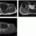
FIGURE 19.6 Neonatal ovarian cyst. A: Coronal T2-weighted fetal MR image performed at 34 weeks’ gestation shows a round, circumscribed, homogeneously hyperintense cyst (arrows) in the right abdomen. The lesion extends from the dome of the bladder (B) to the liver (L). B: Postnatal transverse gray-scale ultrasound image also shows a large simple-appearing cyst (asterisk) extending into the right upper quadrant of the abdomen. A 7 cm simple ovarian cyst was found at surgical exploration, and cystectomy was performed. The right ovary was preserved and demonstrated no evidence of torsion. RK, right kidney.

At ultrasound, a corpus luteum can vary in appearance from an irregular, thick-walled cyst to a more solid appearing, collapsed lesion with low-resistance spectral Doppler waveforms in their wall.36 At contrast-enhanced CT and MRI, the walls of these cysts usually appear thickened and hyperenhancing with crenated margins. The ovulating ovary and corpora lutea also show increased uptake on 18F-FDG-PET (18F-fluorodeoxyglucose positron emission tomography), simulating pelvic lymphadenopathy or ovarian neoplasm.37

Failed ovulation can result in a functional cyst that usually remains simple, but may grow causing pain.36 In postpubertal girls, simple ovarian cysts up to 3 cm most likely represent a dominant follicle (with rupture either impending or failing to occur) or persistence of the corpus luteum as a cyst after ovulation.1,36 Although uncommon, autonomously functioning follicular cysts (Fig. 19.7) are a common cause of peripheral precocious puberty.14 A cyst larger than 9 mm in a girl with precocious puberty should raise a question of an autonomously functioning follicular cyst or hormone-producing tumor14 requiring resection. Observation is generally sufficient for nonfunctioning cysts; however, aspiration may be needed for large ovarian cysts to minimize the risk of future ovarian torsion.14


FIGURE 19.7 This 4.3 cm follicular cyst with luteinization (A) was excised from the ovary of a 6-year-old girl with isosexual precocity. The luteinized cells impart a yellow color to the cyst lining (B). Microscopically, the cyst lining contains follicular cells with the abundant pink and sometimes vacuolated cytoplasm that typifies “luteinized” cells (C) (hematoxylin and eosin, original magnification, 600×).

Hemorrhagic ovarian cysts are generally from corpora lutea, which typically have bloody contents (Fig. 19.8); they vary in appearance depending upon the age of the hemorrhage.1,36,38 At ultrasound, they may appear echogenic without posterior acoustic shadowing; may contain dependent debris, fluid-debris levels, or retracted, avascular, lobulated blood clot (Figs. 19.9 and 19.10); or may contain lacy or cobweb-appearing fibrinous strands. Retracted blood clot within a cyst does not have blood flow on color Doppler ultrasound and may move with ballottement with the transducer (this is evaluated by applying gentle pressure with the transducer followed by release to assess movement within the fluid), whereas a solid tumor nodule does not. The cyst contents should not have
blood flow on Doppler ultrasound evaluation and should not enhance on CT or MRI. At contrast-enhanced CT, a hemorrhagic cyst may appear hyperdense compared to other follicles, but is still less attenuating than surrounding enhanced ovarian stroma. A hematocrit effect may be observed. The MRI appearance depends upon the age of the hemorrhage. Signal hyperintensity on both non-fat-saturated and fat-saturated T1-weighted MR images is a reliable indicator of the presence of hemorrhage. These lesions may show lower than expected signal intensity on T2-weighted MR images, often referred to as “T2 shading,” which is the result of evolving blood products (Fig. 19.10). Other causes of blood-filled ovarian cysts besides corpora lutea include posttorsion/infarct and endometriotic cyst, both rare in childhood.


FIGURE 19.8 Corpus luteum cyst, with a lumen containing a coagulum of blood (hematoxylin and eosin, original magnification, 20×).


FIGURE 19.9 Corpus luteum cyst becoming a hemorrhagic cyst in a 15-year-old girl. A: The corpus luteum cyst (arrow) appears as an anechoic structure with a defined wall and increased through transmission within the right ovary. B: Notice the expected low-resistance spectral Doppler waveform in the adjacent ovarian parenchyma. C: Five days later, the same cyst now contains layering echogenic debris (arrows) and low-level echoes, compatible with a hemorrhagic ovarian cyst.

Functional hemorrhagic cysts typically resolve or at least decrease in size over 1 to 2 menstrual cycles.1 If there is acute and sometimes painful cyst rupture, the cyst may appear collapsed or may be no longer visible with a variable amount of free pelvic fluid that may contain low-level echoes due to hemorrhage. Management is usually conservative. For simple cysts in postmenarchal girls measuring 3 to 5 cm, follow-up is commonly not required.36 Follow-up ultrasound is recommended for simple cysts measuring >5 cm and less than or
equal to 7 cm,36 although aspiration may be warranted to prevent potential torsion. Intervention may also be required for larger cysts for pain control or in case of suspected ovarian torsion, with laparoscopic cyst aspiration or cystectomy most often performed. Simple cysts larger than 7 cm require further evaluation with MR imaging or laparoscopy.36


FIGURE 19.10 A 16-year-old girl with a hemorrhagic left ovarian cyst. A: Axial T2-weighted MR image shows a hemorrhagic cyst (arrow) in the left ovary that demonstrates “T2 shading” phenomenon due to layering blood products within the cyst. This appearance is classically associated with endometriomas, which are typically extraovarian, but can be seen in ovarian hemorrhagic cysts as well. B: Longitudinal color Doppler ultrasound image of the left ovary shows internal avascular retractile blood products within the cyst. The cyst resolved on subsequent follow-up ultrasound evaluation. (Case courtesy of Jonathan R. Dillman, MD, MSc, Cincinnati Children’s Hospital Medical Center, Cincinnati, OH.)


Polycystic Ovarian Syndrome

Polycystic ovarian syndrome (PCOS) consists of hyperandrogenism, enlarged polycystic ovaries, and obesity. Affected patients often present with menstrual cycle irregularity and may have insulin resistance.39 The incidence of PCOS in adolescents is about 3%,40 although 16% of girls presenting with menstrual irregularity were diagnosed with PCOS in one study.41 There is likely a genetic basis for the disease, which has been proposed to represent a form of arrested adrenarche.40 Given the association of this syndrome with insulin resistance and hypertension, implications for long-term cardiovascular health make PCOS important to detect.


FIGURE 19.11 A 16-year-old girl with irregular menstrual cycles, severe acne, and hyperandrogenism. A: Transverse power Doppler ultrasound image of the pelvis shows bilaterally enlarged ovaries (arrowheads) containing multiple subcentimeter cysts. Ovarian volumes measure >15 mL. B: Axial T2-weighted fat-saturated MR image shows an enlarged left ovary with prominent hypointense central stroma (asterisk) and numerous peripherally arranged follicles. These findings can be seen with polycystic ovarian syndrome. Findings are more conspicuous in the left ovary due to the plane of the image.

Diagnostic criteria include physical examination evidence of hyperandrogenism; hormonal imbalances, including testosterone elevation and an elevated LH/FSH ratio; oligo- or amenorrhea; insulin resistance or hyperinsulinemia; and polycystic ovarian morphology (PCOM).40 Accelerated bone maturation may also be present.42 Because ovaries containing multiple follicles of up to 1 cm can be normal during adolescence,43 a diagnosis of PCOS should be only be considered if the abnormal ovarian morphology is accompanied by other clinical or laboratory evidence of PCOS.20,39,40 Normal ovarian morphology does not exclude the diagnosis of PCOS.

The criteria for PCOM include an ovarian volume of more than 10 mL with or without 12 or more follicles in each ovary measuring 2 to 9 mm in diameter44 (Fig. 19.11). Follicular distribution and stromal hyperechogenicity are not included
in these criteria. Although both ovaries are typically affected, findings may be seen in only one ovary.


FIGURE 19.12 A 12-year-old girl with acute pelvic pain. A: Sagittal T2-weighted MR image shows a dilated, fluid-containing right fallopian tube (arrows) with adjacent fluid. B: More lateral MR image shows an adjacent focal fluid collection associated with the right ovary (asterisk). This collection represents a tubo-ovarian abscess. C: Axial-oblique T2-weighted MR image shows the dilated fallopian tube in cross-section appearing as multiple round structures (arrows) as well as the adjacent abscess (asterisk). (Case courtesy of Jonathan R. Dillman, MD, MSc, Cincinnati Children’s Hospital Medical Center, Cincinnati, OH.)

Transvaginal ultrasound is the ideal imaging modality for evaluation of ovaries and ovarian follicles in adolescent pediatric patients with clinically suspected PCOS; however, transvaginal ultrasound may not be appropriate in girls who are not sexually active. When evaluating PCOM with ultrasound, ovarian volumes should be calculated utilizing the simplified formula for a prolate ellipsoid (0.52 × length × width × height).44 If a dominant follicle (>10 mm) or a corpus luteum is encountered, repeat ultrasound is recommended during the next menstrual cycle. Some authors36,45 have suggested a threshold of at least 19 or more follicles as criteria for diagnosing PCOS; however, these studies were performed using transvaginal probes.

Current treatments for PCOS include weight reduction, hair growth suppression and/or removal, insulin-sensitizing agents (e.g., metformin), and hormonal therapy with oral contraceptives or antiandrogens.39,40


Infectious and Inflammatory Disorder


Tubo-Ovarian Abscess

Twenty percent of adolescents with pelvic inflammatory disease (PID) develop a tubo-ovarian abscess (TOA),1 presenting with acute or chronic pelvic pain, often associated with fever and an elevated white blood count. At ultrasound, TOAs are typically complex adnexal collections with variable internal echoes and septations, no internal Doppler blood flow, and a thick, hyperemic wall on color Doppler evaluation.1 TOAs are also well demonstrated by CT and MRI as complex fluid collections with peripheral postcontrast enhancement and adjacent inflammatory changes46 (Fig. 19.12), although ultrasound is diagnostic in many pediatric cases. The ipsilateral ovary may be indistinguishable, and there may be an associated hydro-or pyosalpinx (Fig. 19.13). The differential diagnosis for TOA includes endometrioma, ectopic pregnancy, and malignancy; however, the clinical findings suggesting acute infection are often helpful in arriving at the correct diagnosis. If the patient denies sexual activity, consideration should be given to secondary adnexal involvement due to other disease, such as a ruptured appendix or inflammatory bowel disease.

Management generally requires intravenous antibiotic therapy. For larger, accessible collections, transvaginal or percutaneous image-guided drainage may be an option. Laparoscopic drainage can be performed if needed.


Neoplastic Disorders

The overall incidence of ovarian neoplasms in childhood is low with ovarian malignancies accounting for up to 30% of ovarian masses, comprising 1% of childhood cancers.1,7,47 Twenty-five percent of girls between 1 and 8 years of age presenting with an ovarian mass or precocious puberty have an ovarian malignancy, whereas only 10% of girls ages 9 to 19 years with an ovarian mass have a malignant lesion due to the increased incidence of cysts and benign neoplasms in the older age group.7,48 Ovarian malignancy is very rare under 1 year of age.

The classification of neoplastic ovarian masses is by cell type, including surface epithelial, germ cell, sex cord stromal, metastases, and miscellaneous (Table 19.2). Germ cell tumors comprise 80% of ovarian neoplasms in girls <20 years of age, whereas 95% of ovarian lesions are of epithelial origin in older adults.7,50,51

Typical clinical presentations of children with ovarian neoplasms include pelvic or abdominal mass or precocious puberty.2 Pain due to torsion and/or rupture with hemoperitoneum is also a common presentation,1,52 but less common in children than adults.47 As in adults, solid or heterogeneous appearance of an ovarian mass is concerning for malignancy,2
although 5% of simple ovarian cysts over 6 cm in children may contain malignant cells.7,48 Ovarian lesions over 8 cm are suspicious for malignancy in children.48


FIGURE 19.13 A 17-year-old girl presented with a right-sided pyosalpinx. Transverse ultrasound (A) and coronal T2-weighted fat-saturated MR (B) images through the right lower quadrant show a dilated fallopian tube (arrows) and adjacent inflammation due to pelvic inflammatory disease.

Ultrasound is considered the first-line imaging modality for assessing pediatric pelvic masses and is typically performed using transabdominal technique. Doppler ultrasound evaluation is a helpful adjunct to gray-scale imaging. MRI often allows for additional tissue characterization and helps confirm the organ of origin for indeterminate pelvic lesions.6 CT is generally reserved for staging of disease extension in pediatric patients with ovarian neoplasms, although it also depicts intralesional fat and calcification/ossification.

Management of ovarian tumors in children depends upon the likelihood of malignancy with ovarian-sparing procedures commonly attempted for benign lesions.52,53 A surgical tumor staging workup is performed in cases of suspected malignancy. Additional evaluation includes hormonal assessment in the setting of precocious puberty or virilization. Serum markers, including CA-125, β-hCG (beta-human chorionic gonadotropin), LDH (lactate dehydrogenase), and α-FP (alpha-fetoprotein), are useful at diagnosis as well as at follow-up to monitor response to therapy and relapse.7,48 Adjuvant chemotherapy and/or radiation therapy may also be indicated for some malignant tumors.52,54


Benign Ovarian Neoplasms

Mature teratomas are the only benign germ cell tumor to arise from the ovary,1 and they are the most common ovarian neoplasm in children, accounting for 67%.2 By definition, these lesions must contain at least two of the three germ cell layers—endoderm, mesoderm, and ectoderm.55 These lesions are often called cystic teratomas or dermoid cysts due to their tendency to be mostly cystic and contain dermal elements (Fig. 19.14).1 About 25% are bilateral.2,55

The ultrasound appearance of mature ovarian teratomas is highly variable depending upon the sizes and proportions of cystic, fatty, solid, and calcified components.1 The classic appearance is a predominantly cystic lesion with a solid echogenic mural nodule representing the Rokitansky nodule (or dermoid plug)1,55 (Fig. 19.15). This mural nodule may contain fat, skin, hair follicles, teeth, and/or bone. “Dermoid mesh,” linear echoes representing hair, may be seen.7,36 Echogenic nondependent floating sebum, sometimes creating a fat-fluid level, is another distinguishing feature of mature teratomas at ultrasound. An echogenic focus with prominent posterior acoustic shadowing represents the “tip of the iceberg” sign due to absorption/attenuation of sound waves by hair and fat1,7,36; however, gas within pelvic bowel loops can also create this appearance and is a pitfall. Ballottement with the transducer is a useful technique to elicit motion as a cohesive mass; bowel loops commonly efface.36

At CT, the finding of fat attenuation (<-20 Hounsfield units) in a complex solid or cystic lesion with associated calcification is diagnostic of ovarian teratoma7 (Fig. 19.15); without pathologic examination, it is difficult to exclude the presence of immature areas. At MRI, fat-containing teratomas can be differentiated from hemorrhagic ovarian cysts by comparison of non-fat-saturated and fat-saturated T1-weighted MR images; the fatty elements of an ovarian teratoma typically show signal loss with fat saturation, whereas hemorrhage remains hyperintense (Fig. 19.16). Complications of mature ovarian teratomas include torsion (Figs. 19.17 and 19.18), seen in 15% of cases,1 rupture, malignant transformation, and very rarely autoimmune hemolytic anemia1 and immune-mediated limbic encephalitis.7,56

Only gold members can continue reading. Log In or Register to continue

Stay updated, free articles. Join our Telegram channel

Oct 13, 2018 | Posted by in PEDIATRIC IMAGING | Comments Off on Female Genital Tract

Full access? Get Clinical Tree

Get Clinical Tree app for offline access