
Overview
Anatomy
• The pelvis is that part of the peritoneal cavity extending from the iliac crests superiorly to the pelvic diaphragm inferiorly.
• The female pelvis consists of the genital tract (vagina, uterus, and uterine tubes), ovaries, urinary bladder, a portion of the ureters and intestinal tract, pelvic musculature, ligaments, and peritoneal spaces.
True Pelvis and False Pelvis
The descriptive compartments of the pelvic cavity are the true pelvis and false pelvis:
• The true and false pelves are based on an oblique plane in the pelvis defined by the linea terminalis, an imaginary line drawn from the symphysis pubis around to the sacral promontory, marking the dividing plane.
• The true pelvis (“lesser pelvis” or “pelvis minor”) is the region deep to the linea terminalis.
• The false pelvis (“greater pelvis” or “pelvis major”) is the area superior to the linea terminalis and inferior to the iliac crests.
Pelvic Regions
Right Iliac, Hypogastric, and Left Iliac
The descriptive regions of the pelvic cavity (right iliac, hypogastric, left iliac) are subdivisions of the hypogastrium:
• The right iliac region contains the cecum of the colon, appendix, distal end of the right ureter, and right ovary.
• The hypogastric region includes the distal end of the ileum, urinary bladder, and uterus.
• The left iliac region contains the sigmoid colon, distal end of the left ureter, and left ovary.
Vagina
The vagina is a reproductive organ that is part of the genital tract:
• Located in the midregion of the true pelvis between the urinary bladder (anteriorly) and the rectum (posteriorly), the vagina extends from the external genitalia to the cervix of the uterus.
• The vagina is a muscular, tubular organ. The walls are composed of 3 layers:
1. Inner mucosal lining of epithelial cells
2. Middle thin, smooth muscle wall
3. Outer adventitia
• The linings of the vagina and uterus enclose a continuous cavity or channel through which the fetus passes at birth. The inner epithelium encloses a centrally located vaginal canal that is approximately 9 cm long.
Uterus
Fundus, Corpus, Isthmus, Cervix (Internal and External Os), Endometrium, Myometrium, and Serosa
The uterus is a reproductive organ that is part of the genital tract. It is a muscular, hollow organ where the fertilized ovum embeds and the developing embryo and fetus are nourished:
• The uterus is typically located in the midline of the true pelvis between the urinary bladder (anteriorly) and the rectum (posteriorly). It may also lie just to the right or left of the midline. Its central cavity opens into a uterine (or fallopian) tube on either side and into the vaginal cavity below.
• Uterine walls are composed of 3 layers:
1. Endometrium: inner mucosal layer that encloses the uterine cavity (also called the “endometrial cavity,” “endometrial canal,” or “uterine canal”) that is continuous with vaginal epithelium inferiorly. The thickness of the endometrium varies. Just before the onset of menses it measures 8 mm. Following menses it measures 1 mm. The endometrium consists of 2 layers:
a. Superficial (“functional layer” or “functional zone”): increases in size during the menstrual cycle and partially sloughs off during menses.
b. Deep (“basal layer”): composed of dense stroma and mucosal glands; it is not significantly influenced by the menstrual cycle.
2. Myometrium: middle, smooth muscle layer that forms the bulk of the uterus.
3. Serosa: outer peritoneal layer is a thin membrane that completely covers the myometrium.
• The uterus is pear-shaped and descriptively divided into 4 parts:
1. Fundus: widest and most superior segment that is continuous with the uterine corpus.
2. Corpus or body: largest part of the uterus that is continuous with the uterine cervix.
3. Uterine isthmus: the slightly constricted portion of the uterus where the uterine body meets the uterine cervix.
4. Cervix: lower cylindrical portion of the uterus that projects into the vagina. The cervical portion of the endometrial canal or the endocervical canal extends 2 to 4 cm from its internal os (or opening), at approximately the same level as the ithmus, where it joins the endometrial canal to its external os, which projects into the vaginal canal.
• The size of the uterus is variable and described 4 different ways depending on patient parity and age:
1. Prepubertal size is approximately 2.5 to 3 cm long, 2 cm wide and 1 cm thick. The cervix comprises a significantly greater proportion of the organ.
2. Postpubertal, nulliparous size is usually 7 to 8 cm long, 3 to 5 cm wide, and 3 to 5 cm thick.
3. Multiparous size is usually 8.5 to 9 cm long and 5 cm wide.
4. Postmenopausal size depends on parity but the uterus significantly decreases in size and assumes a prepubertal shape where the cervix comprises the greater portion of the uterus.
• Uterine position can be variable but the uterus normally tilts forward, resting on the dome of the bladder. Due to its peritoneal connections and ligaments, there is considerable mobility of the uterus within the true pelvis that affords minimal displacement of the uterus with the filling of the urinary bladder or rectum, as well as marked displacement of the uterus during pregnancy. Further, the flexibility of the uterine support structures affords considerable variations in normal uterine position that are described in 4 different ways as:
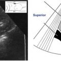
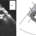
1. Anteverted when the bladder is empty; the vagina and cervix form a 90-degree angle and the body and fundus of the uterus are tilted anteriorly toward the pubic bone. The uterus is typically anteverted (see illustration A on the facing page).
2. Anteflexed when the bladder is empty; the vagina and cervix form a 90-degree angle and the body and fundus of the uterus are bent anteriorly toward the pubic bone until the fundus points inferiorly and rests near the cervix (see illustration B on the facing page).
3. Retroverted when the bladder is empty; the body and fundus of the uterus are tilted posteriorly toward the sacrum until the cervix and vagina are linearly oriented (see illustration C below).

Uterine Tubes (Fallopian Tubes or Oviducts)
Interstitial Segment, Isthmus, Ampulla, and Infundibulum
The uterine tubes are reproductive organs that are part of the genital tract. They are coiled, muscular tubes that emerge from the superolateral margins of the uterus referred to as the uterine cornua, then run laterally within the peritoneum along the superior free margin of the broad ligaments until they reach the ovaries. Uterine tubes are responsible for directing mature ovum (egg cell) from the ovaries to the uterus through gentle peristalsis of its smooth muscle walls:
• Length of the tubes varies from 7 to 12 cm. Their widest diameter is approximately 3 mm.
• Uterine tubes are descriptively divided into 4 segments:
1. Interstitial or intramural is the narrowest segment, which is enclosed by the uterus.
2. Uterine tube isthmus is the segment adjacent to the uterine wall, connected to the interstitial segment. The isthmus is a short, straight, narrow portion of the tube that widens laterally, to form the ampullary and infundiblular segments.
3. Ampulla is the coiled and longest segment where fertilization usually occurs.
4. Infundibulum is the widest and funnel-shaped end of the tube that opens into the peritoneal cavity adjacent to the ovary. Fringelike extensions of the infundibulum called fimbria overlie the ovary and direct the released ovum into the tube.
Ovaries
Ovaries are paired, bilateral, almond-shaped, female reproductive organs in which ova are formed. The ovaries contain numerous follicles, fluid-filled sacs that contain developing eggs (oocytes).
• The ovaries are located in the adnexa (the peritoneal cavity spaces located posterior to the broad ligaments) within the true pelvis. Each ovary is anterior to a ureter and internal iliac artery.
• The position of the ovaries is variable:
• Typically the ovaries lay lateral or posterolateral to the uterus against the pelvic sidewalls when the uterus is characteristically anteverted and lies at the midline of the pelvis.
• The ovaries never move in front of (or anterior to) the uterus or broad ligaments.
• If the uterus is retroverted, the ovaries tend to lay superolateral, adjacent to the fundus of the uterus.
• When the uterus lies to one side of the midline (a normal variation), the ovary on that same side is often displaced from its typical position to lie superior to the fundus of the uterus.
• If the uterus is enlarged, the ovaries are shifted superolaterally.
• After a hysterectomy (surgical removal of the uterus), the ovaries shift toward the midline, superior to the vagina.
• The size of the ovaries varies and is described 3 ways depending on patient age, phase of the menstrual cycle, and menstrual status:
1. Prepubertal ovaries are relatively large at birth, then between the ages of 2 and 6 ovarian size remains relatively stable with a volume of 2.3 cm3 or less. Between the ages of 6 and 7, age-related growth associated with cystic functional changes begin and continue through puberty.
2. Postpubertal ovarian size is approximately 4 to 5 cm long, 3 cm wide, and 2 cm thick. Volume measurements are generally 9.8 cm3 but may be higher during the preovulatory phase and lower during luteal phases.
3. Postmenopausal ovaries are approximately 2 cm long, 1 cm wide, and 0.5 cm thick. Volume measurements are usually 5.8 cm3. It should be noted that ovarian atrophy takes place gradually.
Urinary Bladder
The urinary bladder is a symmetrical, hollow, muscular organ. It is part of the urinary system and serves as a reservoir for the urine that is formed in the kidneys.
• The urinary bladder is fixed inferiorly at its base in the true pelvis, directly posterior to the symphysis pubis and anterior to the uterus and vagina. As the bladder fills with urine, the dome can extend superiorly into the false pelvis.
• Bladder shape is variable depending on distention. The bladder can hold as much as 16 to 18 ounces of urine.
• The normal distended urinary bladder wall measures 1 cm or less.
Ureters
The ureters are muscular tubes that are the part of the urinary system that convey urine from the kidneys to the urinary bladder.
• Normally each kidney has one ureter that travels inferiorly through the retroperitoneum for 10 to 12 inches from the renal hilum to the posterior portion of the bladder. They are less than 1.4 inches wide and decrease in diameter as they course to the bladder.
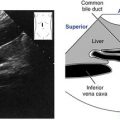
• In the true pelvis the ureter runs between the internal iliac artery (posteriorly) and the ovary (anteriorly), then courses anteromedially to enter the trigone of the urinary bladder just anterior to the vagina.
• Ureters are clinically significant in the female pelvis because pelvic pathologic conditions can cause obstruction of the ureter(s) and ultimately affect the kidney(s).
Colon
Sigmoid and Rectum
The sigmoid colon and rectum are the parts of the gastrointestinal system located within the true pelvis.
• The sigmoid colon is continuous with the descending colon in the left lower quadrant of the pelvis and is loosely secured to the posterior pelvic wall. It descends toward the rectum in the inferoposterior aspect of the pelvis at the level of the third sacral vertebra.
• The rectum is fixed in its position posterior to the vagina and is largely retroperitoneal.
Musculature
Psoas Muscles, and False Pelvis and True Pelvis Musculature
Pelvic musculature is part of the musculoskeletal system. The pelvic muscles serve as support and protection:
• Psoas muscles are the paired major muscles on either side of the spine that extend from the lateral aspects of the lower thoracic vertebrae anterolaterally across the posterior wall of the abdominopelvic cavity to the iliac crests.
• False pelvis musculature: iliopsoas, rectus abdominus, transverse abdominus.
• Each psoas muscle joins an iliacus muscle at the level of the iliac crests to form the iliopsoas muscles that travel anteroinferiorly to insert into the lesser trochanter of the femur.
• The large pair of rectus abdominus muscles extend from the sixth ribs and xiphoid process of the sternum down to the pubic symphysis.
• Each transverse abdominus muscle forms the anterolateral borders of the abdominopelvic cavity. The muscular sheath surrounding each rectus abdominus muscle fuses with the transverse abdominus muscles to form the tendinous linea alba at the midline.
• True pelvis musculature: obturator internus, piriformis, pelvic diaphragm (pubococcygeus, iliococcygeus [levator ani], coccygeus).
• The obturator internus muscles line the lateral walls of the true pelvis.
• Piriformis muscles are situated in the posterior region of the true pelvis behind the uterus. They are often misidentified as enlarged ovaries.
• The pelvic diaphragm is a group of muscles lining the floor of the true pelvis to support the pelvic organs. Each pubococcygeus muscle extends from the pubic bones to the coccyx, encircling the rectum, vagina, and urethra. The iliococcygeus muscles are located lateral to each pubococcygeus muscle. Together these muscles form a hammock across the pelvic floor and are termed the levator ani muscles. Each coccygeus muscle extends from the ischial spine to the sacrum and coccyx. They are the most posterior muscle pair of the pelvic diaphragm.
Ligaments
Broad, Round, Cardinal, Uterosacral, Infundibulopelvic, Ovarian, Pubovesical, and Lateral
The broad ligaments are the only pelvic ligaments routinely identified sonographically. Pelvic ligaments are part of the musculoskeletal system. They are peritoneal connections that provide flexibility and mobility of pelvic organs:
• Broad ligaments: each broad ligament extends between the uterine cornu and ovary. Each uterine (or fallopian) tube, round ligament, ovarian ligament, and vascular structures of the uterus and ovaries are positioned between the 2 layers of the broad ligaments. These structures are surrounded by fat and connective tissue called the parametrium.
Pelvic Spaces
Anterior Cul-de-Sac, Posterior Cul-de-Sac, and Space of Retzius
Differentiation of the 3 pelvic peritoneal spaces is significant because of the potential abnormalities that can invade them:
1. Anterior cul-de-sac, or vesicouterine pouch, is a shallow peritoneal space located between the anterior wall of the uterus and the urinary bladder. This space all but disappears as the urinary bladder fills with urine.
2. Posterior cul-de-sac, rectouterine pouch, or pouch of Douglas is the most posterior and dependent portion of the peritoneal sac. It is located between the rectum and the uterus.
3. Space of Retzius, or prevesical or retropubic space, is a fascial space between the anterior bladder wall and pubic symphysis.
Physiology
Female Reproductive System
Between puberty and menopause, the female reproductive system undergoes monthly cyclical changes referred to as the menstrual cycle. The menstrual cycle usually follows a 28-day course, during which a single ovum reaches maturity and is released into the genital tract. The pituitary gland (part of the brain that oversees all hormonal activity) and ovaries secrete hormones that control changes in the ovaries and the uterine endometrium throughout the menstrual cycle. The changes in the ovaries correlate to the changes in the endometrium.
• Days 1 to 14 of the menstrual cycle correspond to the “follicular phase” of the ovaries and menses and the “proliferative phase” of the uterine endometrium.
• By the onset of menses (menstrual flow or monthly discharge of blood from the uterus of nonpregnant women), each ovary contains thousands of undeveloped follicles, each composed of a single primary oocyte. During the ovarian follicular phase (days 1 to 14 of the menstrual cycle), follicle-stimulating hormone (FSH) is released by the pituitary gland to initiate the development of several primary ovarian follicles. As each primary follicle grows, its oocyte reaches a mature size (ovum). At this stage of development, the ovum and surrounding structures are referred to as secondary ovarian follicles.
• Menses generally occurs during days 1 to 5 of the menstrual cycle. The thickened, superficial layer of the endometrium is shed when fertilization of an ovum does not occur.
• Following menses, the endometrium goes into the proliferative phase that lasts until day 14 of the menstrual cycle. During the proliferative phase, ovarian follicles contain cells that begin to release the hormone estrogen, which initiates thickening and swelling of the endometrium in preparation for implantation by a fertilized ovum.
Related posts:
Stay updated, free articles. Join our Telegram channel

Full access? Get Clinical Tree


