75 Field of View: Rectangular

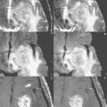
Fig. 75.1
The field of view (FOV) defines the part of the patient to be imaged. This is chosen prior to scan acquisition and need not be square. Indeed, under certain circumstances, a reduced FOV along one axis can be advantageous. The topic of this case is the choice of the FOV in the phase-encoding direction.
In Fig. 75.1, the FOV in the phase-encoding direction (right to left in this instance) was changed from 100% (Fig. 75.1A) to 75% (Fig. 75.1B) to 50% (Fig. 75.1C). A small enhancing left frontal metastasis is illustrated (black arrow, Fig. 75.1A) on postcontrast T1-weighted scans. Images are displayed as acquired, without cropping or differential magnification. Because the pixel size was held constant, fewer phase-encoding steps were required for Fig. 75.1B (three-fourths the number) and Fig. 75.1C (one-half the number). Scan time is directly proportional to the number of phase-encoding steps, and so the scan time of Fig. 75.1B was three-fourths that of Fig. 75.1A
Stay updated, free articles. Join our Telegram channel

Full access? Get Clinical Tree


