| SKULL BASE REGION | Foramen magnum |
| HISTOPATHOLOGY | N/A |
| PRIOR SURGICAL RESECTION | No |
| PERTINENT LABORATORY FINDINGS | N/A |
Case description
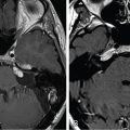
A 55-year-old woman was found to have a right foramen magnum lesion, most likely a meningioma, after undergoing a magnetic resonance imaging (MRI) for new-onset vertigo, later attributed to benign paroxysmal positional vertigo. Serial MRIs studies demonstrated growth at a rate of 1 mm per year ( Figure 11.54.1 ). Because of the small size of the tumor and absence of any symptoms, both radiosurgery and surgical resection were discussed. After careful consideration, the patient opted for stereotactic radiosurgery (SRS) ( Figure 11.54.2 ).
| Radiosurgery Machine | CyberKnife |
| Radiosurgery Dose (Gy) | 15, at the 80% isodose line |
| Number of Fractions | 1 |

Preradiosurgery axial (left) and coronal (right) T1-weighted postgadolinium MRI showing a right foramen magnum mass measuring 1.5 cm.

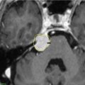
CyberKnife dosimetry plan.
| Critical Structure | Dose Tolerance |
|---|---|
| Brainstem |
|
| Spinal cord | 10–12 Gy |
| Lower cranial nerves | Unknown, but significantly more tolerant than optic nerves |
| Vertebral artery |
|
Related posts:
 Esthesioneuroblastoma – delayed postoperative radiosurgery for recurrence at long-term
 Null cell – delayed postoperative radiosurgery for growing perioptic residual
 Chondrosarcoma – definitive radiosurgery after subtotal resections
 Large vestibular schwannoma – delayed postoperative radiosurgery for growing residual
 Trigeminal neuralgia due to petroclival meningioma – upfront radiosurgery
 Superior sagittal sinus meningioma – delayed postoperative, multisession radiosurgery for growing residual
Stay updated, free articles. Join our Telegram channel

Full access? Get Clinical Tree


