Foramen magnum/petroclival meningioma – immediate postoperative radiosurgery for residual
SKULL BASE REGION
Foramen magnum
HISTOPATHOLOGY
Meningioma, WHO grade 1
PRIOR SURGICAL RESECTION
Yes
PERTINENT LABORATORY FINDINGS
N/A
Case description
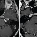
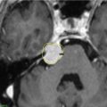
A 63-year-old woman presented with a worsening headache and was found to have a 4-cm petroclival meningioma centered on the lower third of the clivus with compression of the brainstem and a 360-degree encasement of the right vertebral artery ( Figure 11.55.1 ). The patient underwent subtotal resection through a suboccipital retrosigmoid approach with an estimated 90% tumor resection. Residual tumor was left at the foramen magnum, where the vertebral artery was encased by the tumor. Postoperatively, she developed a right hypoglossal nerve palsy. Follow-up imaging demonstrated rapid regrowth of the tumor residual at the foramen magnum, with recurrence of brainstem compression ( Figure 11.55.2 ). This recurrent tumor was resected through a far lateral approach 29 months after the initial operation. The surgical goal of brainstem decompression was achieved with residual tumor left on the foramen magnum and vertebral artery ( Figure 11.55.3 ). She then underwent stereotactic radiosurgery (SRS) of the residual tumor about 3 months after surgery ( Figure 11.55.4 ).
Radiosurgery Machine
CyberKnife
Radiosurgery Dose (Gy)
30, at the 80% isodose line
Number of Fractions
5
Figure 11.55.1.
Axial (left) and coronal (right) T1-weighted postgadolinium MRI before initial surgery.
Figure 11.55.2.
Axial (left) and coronal (right) T1-weighted postgadolinium MRI before the second surgery.
Only gold members can continue reading. Log In or Register to continue
Apr 6, 2024 | Posted by drzezo in GENERAL RADIOLOGY | Comments Off on Foramen magnum/petroclival meningioma – immediate postoperative radiosurgery for residual


 Esthesioneuroblastoma – delayed postoperative radiosurgery for recurrence at long-term
 Null cell – delayed postoperative radiosurgery for growing perioptic residual
 Chondrosarcoma – definitive radiosurgery after subtotal resections
 Large vestibular schwannoma – delayed postoperative radiosurgery for growing residual
 Trigeminal neuralgia due to petroclival meningioma – upfront radiosurgery
 Superior sagittal sinus meningioma – delayed postoperative, multisession radiosurgery for growing residual


