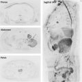
With recent advances in MR imaging, its application in the thorax has been feasible. The performance of both morphologic and functional techniques in the evaluation of thoracic malignances has improved not only differentiation from benign etiologies but also treatment monitoring based on a multiparametric approach. Several MR imaging–derived parameters have been described as potential biomarkers linked with prognosis and survival. Therefore, an integral approach with a nonradiating and noninvasive technique could be an optimal alternative for evaluating those patients.
Key points
- •
Functional MR imaging is an emerging technique in the thorax providing an integral assessment of several characteristics of chest neoplasms by means of prognostic and reproducible quantitative parameters.
- •
Functional MR imaging represents an alternative to fludeoxyglucose F 18 ( 18 FDG)-PET/CT in the differentiation of benign and malignant entities, allowing the accurate staging and treatment response monitoring.
- •
Although there is only initial experience, the performance of functional MR imaging in the heart and chest wall can enhance its utility in the thorax.
Introduction
Several tumor characteristics may be assessed using functional imaging techniques ( Table 1 ). Currently, the most widely accepted tumor metabolism imaging is performed with 18 FDG-PET/CT.
| Technique | Biophysical Basis | Quantitative Parameters |
|---|---|---|
| DWI–MR imaging | Cellularity and tortuosity of interstitial space | ADC |
| DKI | Tissue microstructure | D app ; K app |
| IVIM | Blood flow and tissue diffusivity | f, D, D* |
| DCE–MR imaging | Blood flow and permeability | K trans , V e , V p , K ep , AUC |
| BOLD | Hypoxia | Tissue R2 ∗ relaxivity |
| ASL | Blood flow | Flow quantification |
| Spectroscopy | Metabolism | Ratio of choline to other metabolites |
Although the application of thoracic MR imaging is technically demanding, its great advantage is the possibility of integrating several morphologic and functional techniques in the evaluation of different tumor characteristics. From these techniques, several quantitative and reproducible parameters can be calculated, some of which serve as widely accepted prognostic biomarkers. This article reviews currently available techniques and some of the more promising potential applications of thoracic functional MR imaging.
Functional MR imaging in the chest: technical considerations
Diffusion-Weighted Imaging
Diffusion-weighted imaging (DWI) focuses on the evaluation of brownian motion of water motion in biologic tissues, which has been related to tissue cellularity and architecture (see Table 1 ; Table 2 ). Thoracic application is technically demanding, requiring the application of echo planar imaging (EPI) readout and parallel acquisition strategy. The main hurdle is the presence of motion artifacts and geometric distortion secondary to B0 inhomogeneities and accumulation of phase error during echo train length.
| Signal Intensity High b Value | Signal Intensity ADC Map | Interpretation |
|---|---|---|
| Hypercellular tumors. Rarely, liquid or viscous abscess or blood products | ||
| T2 shine-through; liquefactive necrosis | ||
| Fluid, necrosis, low cellularity lesions, well-differentiated adenocarcinomas | ||
| Fibromuscular tissues, fat, susceptibility artifacts (T2 dark-through effect) | ||
| Mature fibrosis with lower water content |
Motion compensation techniques are often necessary to improve image quality. Respiratory triggering is usually preferred to a single breath hold. For avoiding pulsation artifacts, cardiac triggering (which increases acquisition time) may be useful, especially for the assessment of small lesions located near the heart ( Table 3 ).
| Techniques | Sequence Type/Parallel Accelerating Factor | B Values (s/mm 2 ) | TR/TE (ms) | Resolution (mm 3 ) | Synchronization | Fat Suppression Technique | Image Evaluation | Quantitative Parameter | Acquisition Time |
|---|---|---|---|---|---|---|---|---|---|
| DWI – 3T (IVIM + DKI) | SS EPI/factor 2 | 0, 50, 100, 500, 1000, 1500 | 5000/55 | 2.5 × 2.5 × 7 | Respiratory triggered | Spectral fat suppression | Monoexponential, IVIM, and DKI model | ADC D, D*, f D app ; K app | 4 min 41 s |
| DWI – 1.5T (IVIM + DKI) | SS EPI/factor 2 | 0, 50, 100, 500, 1000, 1500 | 1400/100 | 3 × 3 × 7 | Respiratory triggered | Spectral fat suppression | Monoexponential, IVIM and DKI model | ADC D, D*, f D app ; K app | 5 min 48 s |
| DWI cardiac – 3T | SS EPI/factor 2 | 0, 50, 150, 300 | 1000/44 | 2.58 × 2.54 × 10 | Breath hold; ECG based in diastole | Spectral fat suppression | Qualitative/semiquantitative | ADC ADC ratio SI ratio | 1 min 45 s |
| DWI cardiac – 1.5T | SS EPI/factor 2 | 0, 50, 300 | 2250/96 | 2.6 × 2.54 × 8 | Breath hold; ECG based in diastole | Spectral fat suppression | Qualitative/semiquantitative | ADC ADC ratio SI ratio | 2 min 25 s |
Apparent diffusion coefficient (ADC) represents the exponential signal decay of water molecules on a voxel by voxel basis. Although ADC may be calculated using only 2 b values, the more b values included in its measurement, the more accurate it is.
Recently, advanced models of DWI have been proposed to better understand the behavior of water motion in the different tissues. The intravoxel incoherent motion (IVIM) model of diffusion signal decay has been shown to better fit than monoexponential analysis, especially in the evaluation of well vascularized organs. First, diffusion decay signal shows a rapid attenuation at low b values (b<100–150 s/mm 2 ) secondary to bulk motion of water molecules in capillaries (perfusion effects on diffusion). With higher b values (more than 100 s/mm 2 ), a slower decay of signal occurs due to the real diffusion of water molecules ( Fig. 1 ). IVIM-derived parameters are, theoretically, more reliable markers of tissue diffusivity than ADC and can separate both compartments of diffusion signal decay ( Box 1 ). In addition, ADC assumes a gaussian diffusion behavior, which does not always exactly fit to the real signal decay of diffusion signal. Diffusional kurtosis imaging (DKI) quantifies the deviation of tissue diffusion from a gaussian pattern by measuring diffusion with ultrahigh b values greater than 1500 s/mm 2 (see Fig. 1 ). IVIM and DKI models have been recently explored in the evaluation of chest malignancies and show some promise over conventional ADC measurements.
IVIM-derived parameters
True diffusion of tissue H 2 O molecules (D): not influenced by movement of water molecules within the capillaries
Perfusion contribution to diffusion signal (f): fractional volume of flowing water molecules within the capillaries.
Perfusion contribution to signal decay (D*): amount of nondiffusional random movements of water molecules
DKI-derived parameters
D app estimation of the diffusion coefficient in the direction parallel to the orientation of diffusion sensitizing gradients.
Apparent diffusional kurtosis (K app ): measures the deviation of the true diffusion from a gaussian pattern
Dynamic Contrast-Enhanced–MR Imaging
Dynamic contrast-enhanced (DCE)–MR imaging provides information of tumor physiology, including blood flow, vascular volume, and permeability. DCE–MR imaging also provides imaging insight pertinent to tumor angiogenesis.
Free breathing, high temporal resolution, 3-D gradient-echo sequences are usually acquired during a 5-minute period of time. They have limited coverage and require the use of parallel imaging techniques. Motion and respiratory artifacts are common problems that require correction software during postprocessing.
Different strategies for analysis and quantification of DCE–MR imaging have been reported. The easiest and most used approach is to evaluate the variation of contrast enhancement during multiple time points of the lesion, obtaining time intensity curves (TICs). In the graphic representation of contrast dynamics, the first half is correlated with tumor angiogenesis and the second half with tumor interstitium. This approach does not separate perfusion and permeability. There are several TIC-derived descriptors that can be used for assessing perfusion ( Box 2 ).
TIC-derived parameters
Contrast arrival time (s): time when contrast arrives to a pixel
Time to peak (s): time to maximum SI
Maximum Relative Signal Enhancement (RSE): maximum SI value related to initial non–contrast-enhanced intensity
Wash-in: represents the velocity of enhancement
Washout (WO): represents the velocity of enhancement loss
Initial area under the curve (IAUC): integral of the contrast concentration time curve for a specific time interval. It accounts for blood flow and motion, permeability, extracellular space, and microvessel density.
Quantitative derived parameters from a pharmacokinetic (compartmental) model
K trans (min −1 ): transfer rate of blood from plasma space to extracellular space (transfer constant)
K ep (min −1 ): transfer rate of blood from extracellular space to plasma space (elimination constant)
V e : volume of extracellular space inside a voxel
V p : volume of plasma space inside a voxel
Once the contrast concentration is derived from each dynamic time point acquisition, the tissue status can be quantitatively assessed by calculating the contrast concentrations in the tissue and feeding artery. After applying a contrast kinetic model, quantitative hemodynamic parameters may be obtained. These are based on the use of compartments, defined as spaces where the contrast is evenly distributed. Two well-differentiated models are commonly used, monocompartmental and bicompartmental , according to if only the vascular volume (P) or also the extracellular compartment (E) is considered (see Box 2 ).
The bicompartimental model assumes that: P and E are compartments; E does not exchange tracer with the environment; the clearances for the outlets connecting P and E are equal and the clearance for the outlet of P to the environment equals the plasma flow. In some situations, a tissue with this configuration is reduced to a monocompartmental model:
- 1.
When one of the spaces has a negligible volume
- 2.
When the tracer extravasates with a slow rate (very low concentration at E)
- 3.
When the tracer extravasates with a high rate (single well-mixed space)
Compartmental models are more technically demanding and more time consuming but have demonstrated usefulness in monitoring treatment response and recurrence, because they provide reproducible measurements. Depending on which parametric approximation is used, derived parameters differ in their interpretation (see Box 2 ). Depending on whether or not the delivery of the contrast medium to the tissue is sufficient, K trans approximates the permeability surface area product or blood flow, respectively. Lack of standardization of different models and sequence designs have prevented them from being introduced in the clinical setting.
Functional MR imaging of the lung
Worldwide, lung cancer is the leading cause of cancer-related deaths. CT usually constitutes the first modality in evaluation and staging. It is often complemented with 18 FDG-PET/CT. Other functional modalities have emerged to increase the accuracy of detection, staging, and treatment monitoring. Morphologic MR was classically set as a second-line diagnostic procedure in the evaluation of patients with lung cancer. Recent improvements in hardware and functional techniques, however, have positioned chest MR as an alternative to 18 FDG-PET/CT.
Pulmonary Nodule Detection and Characterization
Functional MR imaging and pulmonary nodule detection
One of the most common challenges in pulmonary medicine is the detection and characterization of pulmonary nodules (PNs). CT is considered the best imaging technique for PN detection.
DWI has been shown useful in the management of PNs. DWI may detect 86.5% of nodules between 6 mm and 9 mm and 97% of PNs greater than 10 mm. In lesions less than 5 mm, the accuracy of DWI falls to only 43.8%, below the sensitivities of other MR imaging sequences, such as short-tau inversion recovery (STIR) (81.5% for lesions >3 mm).
Diffusion-weighted imaging and pulmonary nodule characterization
DWI has shown great usefulness in differentiating benign from malignant PNs ( Table 4 ). With an accuracy cited at 91%, it is comparable to other functional techniques for this differentiation (93%, 94%, and 94% for DCE-CT, 18 FDG-PET/CT, and DCE–MR imaging, respectively). DWI has similar limitations compared with PET in lesion characterization but with lower false-positive results ( Fig. 2 ; Table 5 ).
| Region/Lesion Evaluation | MR Field Strength | B Values | Threshold | Sensitivity/Specificity/Accuracy | |
|---|---|---|---|---|---|
| PN characterization (ADC) | 1.5T and 3T | 0, 500, b1000 s/mm 2 | 1.1–1.4 × 10 −3 mm 2 /s | Pooled 83%/81%/91% | Sensitivity (70%–90%)/specificity (74%–100%) |
| LN characterization (ADC) | 1.5T | 0, 300, 600 s/mm 2 | 1.85 × 10 −3 mm 2 /s | 96.4%/71.4%/83.9% | Pooled sensitivity, specificity, and accuracy: 77.4%–80%, 89%–95%, and 84.4%–97%, respectively Described PPV and NPV: 95.2% and 77.1%, respectively |
| Pleural lesion (ADC) | 3T | 0, 50, 100, 500, 750, 1000 s/mm 2 | 1.52 × 10 −3 mm 2 /s | 71.4%/100%/87.1% | Differentiation of benign versus malignant |
| Mediastinal mass (ADC) | 1.5T | 0, 300, 600 s/mm 2 | 1.56 × 10 −3 mm 2 /s | 95%/96%/94% | Exclusion of cystic mediastinal masses PPV and NPV: 94% and 96%, respectively |
| Cystic mediastinal mass (ADC) | 3T | 0, 100, 900 s/mm 2 | 2.5 × 10 −3 mm 2 /s | 100%/100%/84% | With this cut-off, the ADCs of all cystic benign lesions were higher and the ADCs of malignant ones lower. |
| Diagnostic Modality | False-Positive Results | False-Negative Results | |
|---|---|---|---|
| 18 FDG-PET/CT | PNs | Inflammatory nodules | Well-differentiated adenocarcinoma Small PNs and metastasis |
| LNs | Inflammatory LNs | Small intranodal tumor deposits | |
| Pleural lesions | Inflammatory pleuritis Talc pleurodesis Tuberculous plaques Parapneumonic effusion | Well-differentiated tumors | |
| DWI | PNs | Intranodular inflammation | Mucinous content (T2 shine through) Intranodular necrosis Nonsolid; part-solid lesions (high b value) Low-grade adenocarcinoma Small PNs and metastasis |
| LNs | Tuberculous and nontuberculous granulomatous inflammation | Small intranodal tumor deposits | |
| Pleural lesions | Fibrous plaques (T2 dark-through) | Intratumor necrosis and inflammation | |
Varying results among quantitative characterization of PNs with DWI between series may be explained by the lack of standardization of acquisition and postprocess evaluation ( Figs. 3 and 4 ). Koyama and colleagues reported significant differences in minimum and mean ADC in the evaluation of lung tumors, reflecting the inherent heterogeneity of these lesions. Usuda and colleagues applied a DWI signal intensity (SI)-based analysis revealing that DWI SI was an indicator of the amount of tumor cells and its distribution. Tumor SI of the lesion-to–spinal cord ratio (LSR) of malignant lesions was found higher compared with that of benign ones. Contrarily, Liu and colleagues did not obtain any significant differences of the LSR between malignant and benign nodules. Koyama and coworkers, however, found significantly higher specificity and accuracy of LSR compared with ADC and IVIM-derived parameters in the differentiation of benign and malignant pulmonary nodules. They did not find significant differences in ADC, diffusion coefficient (D), and perfusion fraction (f) between malignant and benign PNs (see Figs. 3 and 4 ).


