Multiparametric MR imaging (mpMRI) combine different sequences that, properly tailored, can provide qualitative and quantitative information about the tumor microenvironment beyond traditional tumor size measures and/or morphologic assessments. This article focuses on mpMRI in the evaluation of urogenital tract malignancies by first reviewing technical aspects and then discussing its potential clinical role. This includes insight into histologic subtyping and grading of renal cell carcinoma and assessment of tumor response to targeted therapies. The clinical utility of mpMRI in the staging and grading of ureteral and bladder tumors is presented. Finally, the evolving role of mpMRI in prostate cancer is discussed.
Key points
- •
Multiparametric MR imaging (mpMRI) protocols include standard sequences tailored for the morphologic evaluation of urogenital tract malignancies that take into account specific needs, such as spatial resolution, respiratory compensation strategies versus breath-hold imaging, and anatomic coverage.
- •
mpMRI also includes acquisitions that provide information about the tumor microenvironment and that extend beyond their morphologic assessment, such as diffusion-weighted imaging (DWI), dynamic contrast-enhanced (DCE) MR imaging, and arterial spin-labeled (ASL) strategies.
- •
mpMRI may offer detailed preoperative insight into renal cell carcinoma (RCC) histologic subtype and grade and provide an opportunity to quantitatively assess tumor response to targeted therapies in patients with metastatic disease.
- •
DWI and apparent diffusion coefficient (ADC) values may play a role in predicting histologic grade and potential treatment response of urothelial carcinoma.
- •
The role of mpMRI in the evaluation of prostate cancer extends beyond tumor staging and now includes disease identification/localization prior to targeted biopsy as well as follow-up of patients on active surveillance.
Introduction
The term, mpMRI , is increasingly used in reference to an approach that takes advantage of the added value of different MR imaging acquisitions to evaluate patients with different tumors, including genitourinary (GU) malignancies. These approaches often include anatomic T1-weighted and T2-weighted images of the region of interest combined with other acquisitions, such as DWI and/or DCE imaging, to provide information about the tumor microenvironment beyond what can be achieved with any single sequence alone. Appropriately performed, these mpMRI protocols offer anatomic insight and possibly qualitative, semiquantitative, and fully quantitative imaging biomarkers, which attempt to reflect the underlying tumor histopathology and biological behavior. This is best illustrated in the characterization and risk stratification of renal and prostate masses, grading of ureteral malignancies, and staging of bladder cancer, settings where the radiologist has the opportunity to influence clinical management. Moreover, these methods can be applied to quantitatively monitoring tumor response to therapy.
This review discusses technical aspects and the clinical role of mpMRI protocols in the evaluation of malignancies of the urogenital tract, including the kidney, ureter, bladder, and prostate.
Multiparametric MR imaging—techniques
Most mpMRI protocols share similar imaging strategies, although different sequences may be applied depending on the body part imaged due to anatomic and practical considerations. Imaging the kidneys has inherent challenges resulting from large degree of motion associated with respiration compared with imaging the prostate, for example, which does not move as much. The size of these organs also influences the spatial resolution of the mpMRI acquisitions. Thus, fast, motion-insensitive sequences or motion-compensated strategies with modest spatial resolution are preferred in the abdomen compared with longer, high-resolution scans in the pelvis.
The MR imaging protocols used for the evaluation of renal masses have been discussed elsewhere. Examinations can be performed at either 1.5T or 3T, although use of a phased-array body coil is mandatory for an optimal examination. Both 2-D and 3-D acquisitions are used in most renal mass protocols.
Although standard MR imaging protocols for abdominal imaging allow for evaluation of the ureters, a dedicated magnetic resonance (MR) urography (MRU) protocol offers advantages in the assessment of primary and secondary malignancies involving the ureters. The MRU protocol at the authors’ institution is similar to the mpMRI protocol for evaluation of renal masses with the addition of several sequences to optimize visualization of the collecting systems, ureter, and bladder (discussed later).
Dedicated mpMRI protocols for assessment of bladder tumors are less broadly adopted in clinical practice, although they are described elsewhere. Optimal evaluation requires high spatial resolution images with a phased-array surface coil. Dedicated pelvic coils or those with larger number of coil elements over a small anatomic area, such as a cardiac coil, tend to provide better image quality for targeted imaging of the pelvis. The use of an endorectal coil (ERC) for evaluation of bladder base and posterior bladder tumors has also been described elsewhere, although it is not routinely used at the authors’ institution.
There are a few unique issues that must be considered when imaging the bladder. Small tumors may be obscured in an underdistended bladder. Bladder overdistention may lead to discomfort, which might result in increased voluntary motion and decreased sensitivity to flat and small tumors. Optimal bladder distention can be achieved by asking the patient to void 2 hours prior to imaging. Assessment of small lesions before and after contrast can be further challenged by progressive filling of the bladder during the MR imaging examination, making the image coregistration difficult. Motion artifacts as a result of bowel peristalsis can degrade images, although administering hyoscyamine (oral or sublingual) or glucagon (intramuscular or intravenous) prior to imaging can be helpful. Finally, chemical shift artifacts at the fat-water interface are commonly encountered when imaging the bladder and can limit evaluation of the bladder wall. Increasing the receiving bandwidth and changing the direction of the frequency-encoding gradient are ways to reduce or displace this artifact, respectively.
The standard sequences in the mpMRI protocol for the evaluation of the prostate have been described elsewhere and are now widely accepted. Currently, there is, however, no consensus regarding the appropriate choice of hardware when imaging the prostate, specifically regarding whether an ERC should be used. At 1.5T, the use of an ERC is generally supported and is superior to an external coil alone for evaluation of prostate cancer. 3T imaging without an ERC is supported by the superior signal-to-noise ratio (SNR) compared with 1.5T scanners, as well as lower costs, improved patient work-flow, and presumed patient acceptance due to discomfort associated with the ERC. The reported image quality of 3T MR imaging without ERC is similar that of 1.5T MR imaging using an ERC ; however, comparisons of diagnostic performance with both 1.5T and 3T strategies with and without the ERC are lacking.
Prostate imaging is optimized by adequate patient preparation. Patients are asked to avoid ejaculation at least 3 days prior to imaging to increase seminal vesicle distention. In postbiopsy patients, imaging should be avoided, if possible, for at least 6 weeks after the procedure because hemorrhage and architectural distortion may mimic or obscure tumors as well as capsular disruption.
T2-Weighted Sequences
Depending on the organ of interest, T2-weighted spin-echo acquisitions can be obtained using either multishot or single-shot techniques, with or without fat suppression. The authors prefer half-Fourier single-shot turbo spin-echo (HASTE)/single-shot fast spin-echo (SSFSE)/single-shot turbo spin-echo (SSTSE) sequences for T2-weighted imaging of the kidneys and upper GU tract. The fast, sequential acquisitions enabled by these sequences are particularly useful in the upper abdomen where diaphragmatic motion can result in significant image degradation. The addition of fat-suppression strategies helps improve image contrast.
T2-weighted multishot sequences provide the additional benefits of greater image contrast, SNR, and spatial resolution compared with single-shot techniques, although they are more vulnerable to motion artifacts. Multishot techniques can be combined with a variety of respiratory compensation strategies to eliminate or improve motion artifacts, although imaging times are further increased. Novel acquisition strategies, such as those using rotating blades during k -space acquisition (eg, Propeller [GE Healthcare, Waukesha, WI], Blade [Siemens Healthcare, Erlangen, Germany], MultiVane [Philips Healthcare, Best, The Netherlands]), are particularly effective in eliminating respiratory-associated artifacts.
Heavily T2-weighted images using long echo times and thick-slice profile acquired in the coronal plane are useful in the evaluation of the collecting system and ureters. These rely on the intrinsic long T2 relaxation time of urine to display hyperintense signal in the GU track against the dark, suppressed background on a single 2-D image. Evaluation of the GU tract is feasible using a variety of 2-D breath-held, thick-slab acquisitions, such as HASTE/SSFSE/SSTSE or rapid acquisition with relaxation enhancement, as well as respiratory-triggered 3-D fast spin-echo (FSE)/turbo spin-echo sequences. Fat saturation techniques can provide additional optimization in the background suppression. Volumetric acquisitions help evaluate complex anatomy and pathology using multiplanar reconstructions and 3-D reformations, such as maximum intensity projection and volume rendering. In contrast, the short acquisition time (ie, approximately 2 seconds per image) of 2-D acquisitions offer the opportunity to create cinelike acquisitions, which can be helpful in distinguish areas of ureteral peristalsis from true pathology (ie, stenosis).
Multiplanar high-resolution, free-breathing, T2-weighted multishot FSE images are preferred for evaluation of malignancies in the bladder. A saturation band placed over the subcutaneous fat in the anterior abdominal wall can help mitigate respiratory ghosting artifacts, although caution must be taken to avoid obscuring the anterior bladder. Other respiratory compensation strategies and alternative k -space acquisitions, such as rotating blades in k -space, may be useful although usually require longer acquisition times. Patient motion artifacts can still be problematic in some cases and may necessitate use of motion-insensitive SSFSE techniques. Steady-state free-precession sequences provide images with very high SNR where fluid and blood vessels are hyperintense, although limited by off-resonance artifacts when applied to a large field of view. The addition of fluid-attenuated inversion recovery strategies has been advocated to achieve T2-weighted evaluation of tumors while suppressing hyperintense urine signal.
Assessment of patients with known or suspected prostatic cancer requires high-resolution, small field of view (eg, 180–220 mm) multiplanar T2-weighted FSE images, with axial images considered the workhorse for depiction of anatomy with optimal distinction between the intermediate-to-high signal intensity of the peripheral zone and the heterogeneous, high and low signal intensity of the central gland in most patients. These images are also the most important to assess extraprostatic extension in most patients. Coronal and sagittal images can be included to help distinguish nodular features of benign prostatic hypertrophy (BPH) from ill-defined tumors as well as wedge-shaped abnormalities in prostatitis. These can also help in the assessment of areas suspicious for extraprostatic or seminal vesicle invasion. An FSE acquisition with an echo time between 90 to 120 ms provides optimal soft-tissue contrast for delineation of prostatic cancers against the hyperintense peripheral zone. Detection of extraprostatic extension may be improved with the utilization of a 3-D FSE acquisition with isotropic voxel resolution. A quantitative approach has been recently proposed with a whole-gland T2 mapping using a multiecho T2-weighted FSE sequence in approximately 6 minutes. Although the role of this type of acquisition has not been yet established, it provides an opportunity to explore additional quantitative measures of tumor characteristics in the context of mpMRI of the prostate.
T1-Weighted Sequences
When imaging the abdomen, the use of gradient-recalled echo (GRE) techniques for the acquisition of T1-weighted images provides rapid imaging (ie, full renal coverage in 15–20 seconds) with an adequate SNR. Depending on the selection of imaging parameters, these images can serve a variety of roles, including, but not limited to, the detection of lipids and noncontrast assessment for vessel patency.
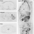
2-D GRE T1-weighted in-phase (IP) and opposed-phase (OP) images, or chemical shift imaging, of the abdomen are used in the renal mass and MRU protocols for the detection of intravoxel lipids. Recently developed 3-D dual-echo Dixon-based acquisitions allow for acquisition of thinner, contiguous slices in a single breath-hold that can then be reconstructed into water, fat, IP, and OP imaging data sets. The thinner slices allow for detection of smaller amounts of intratumoral lipids. Alternatively, Dixon-based sequences with multiple echoes (eg, 6 echoes) offer a way to not only detect but also quantify intralesional lipids, which may provide additional information about tumor biology ( Fig. 1 ).
Determination of the tissue T1-relaxation time (T1-mapping) may be necessary when performing quantitative analysis of DCE data sets. There are different strategies to accomplish this, such as separate T1 acquisitions obtained with the same parameters as those for the DCE protocol but with variable flip angles (eg, 2°, 5°, and 10°), Look-Locker acquisitions, and modified Look-Locker acquisitions.
Contrast-Enhanced Imaging
Although the terms, DCE MR imaging and multiphasic contrast-enhanced (MCE) MR imaging , are often used interchangeably, these refer to different imaging acquisition strategies. MCE MR imaging and CT are analogous in that images are acquired during specific phases, usually at 2 to 4 specific times after the contrast administration. MCE MR imaging protocols are based on lower temporal, higher spatial-resolution acquisitions, which provide some level of temporal information while maintaining the high spatial resolution needed for characterization of disease and treatment planning in clinical practice. This method is best typified by renal imaging (ie, corticomedullary, nephrographic, and excretory phases) and is used to provide a qualitative assessment of enhancement characteristics of a specific organ and/or pathophysiologic condition.
DCE MR imaging involves a lower spatial, higher temporal resolution strategy during the acquisition of multiple serial images before, during, and after intravenous injection of gadolinium to provide a more detailed, quantitative, and/or semiquantitative pharmacokinetic assessment of enhancement. This technique is most often used in the evaluation of prostate masses and, to a lesser extent, renal masses and bladder tumors. Accurate quantitative assessment of enhancement characteristics in a tumor usually requires high temporal resolution imaging, because it allows for the acquisition of a greater number of images. This may be dependent, however, on the intrinsic pathophysiologic characteristics of the tumor. For example, the vascularity in the prostate and in prostate cancer is low compared with that of the kidneys and renal cancer, respectively. Thus, a higher temporal resolution may be necessary for assessment of renal masses than that of prostate cancer. MCE MR imaging with high spatial resolution offers a semiquantitative, although potentially less accurate, description of enhancement characteristics.
Kidney
At the authors’ institution, 3-D fat-suppressed spoiled gradient echo (SPGR) T1-weighted sequences are acquired in the coronal plane. The water reconstruction of Dixon-based acquisitions is preferred because of the more robust, homogeneous fat suppression compared with frequency-selective fat saturation strategies, particularly at high field strength (ie, 3T). A semiquantitative assessment of renal mass enhancement is accomplished by an acquisition during the corticomedullary phase (timed to the arrival of contrast to the kidneys with an MR imaging fluoroscopic technique) followed by images obtained during the early (ie, 40 s) and late (ie, 90 s) nephrographic phase. Sagittal oblique images of each kidney and axial images are obtained during the excretory phase.
A more quantitative assessment of vascularity in renal masses is feasible using a DCE MR imaging protocol, although respiratory motion and the needs for anatomic coverage may dictate the acquisition strategy. An acquisition of a single slice through the renal mass with a saturation prepared 2-D T1-weighted acquisition provides a motion-insensitive (ie, free breathing) and a very fast (<2 s) temporal resolution ; however, this approach provides limited tumor coverage. Alternatively, a 3-D acquisition with a SPGR sequence provides an assessment of the entire tumor although with lower temporal resolution and, thus, generally incompatible with free-breathing acquisitions. The authors prefer the whole-tumor assessment using a 3-D SPGR acquisition with a 5-second temporal resolution acquired in groups of 3 sets of images within a 15-second breath-hold. These are alternated with 15-second periods for breathing over a 6-minute total acquisition time. Newer acquisitions using alternative k -space filling approaches, such as different versions of k -hole, radial, spiral, and view-sharing strategies, may provide the opportunity to obtain high-quality, free-breathing, whole-tumor DCE data sets.
Ureter
Neoplasms arising from or involving the ureters are usually evaluated with an MCE MR imaging protocol and qualitative assessment of enhancement characteristics. The authors use an magnetic resonance urography (MRU) protocol, which comprises the same MCE acquisitions as that of the renal mass protocol, with the addition of an excretory phase acquisition, which is typically obtained 5 minutes after intravenous contrast administration. When using a standard dose of contrast, concentrated gadolinium in urine results in dark signal intensity due to T2* effects. The intravenous administration of 5 to 10 mg of furosemide prior to injection of gadolinium facilitates the evaluation of the collecting system by increasing water excretion and distention of the collecting system as well as dilution of the concentrated gadolinium, which becomes hyperintense. Although administration of a low-dose of gadolinium has also been suggested, MRU in the absence of a pharmacologic diuresis is limited by lack of distention of the collecting system.
Bladder
The use of DCE MR imaging or MCE MR imaging approaches for assessment of bladder tumors varies among institutions, because the utility remains controversial. MCE MR imaging acquisitions may provide a better delineation of the tumor extension in some patients, although local staging is usually based on high-resolution T2-weighted images. A single-slice, high-temporal resolution DCE protocol has been proposed for the characterization and staging of bladder cancer. This approach is not widely used, however, in clinical practice, mainly due to limited anatomic coverage to assess the entire tumor. Again, the newly developed 3-D T1-weighted acquisitions may offer an opportunity to assess the whole-tumor with quantitative DCE MR imaging, although the experience with these techniques is still limited.
Prostate
A detailed discussion of DCE imaging of the prostate and the various means of analysis (ie, kinetic compartmental modeling) is beyond the scope of this review. DCE MR imaging of the prostate is accomplished with a 3-D T1-weighted SPGR sequence covering the entire prostate and acquired before, during, and after the administration of a single dose of a gadolinium-based contrast agent administered at 2 to 4 mL/s followed by a 20-mL saline flush. Images are acquired repeatedly for at least 5 minutes. There is debate about the optimal temporal resolution of DCE MR imaging acquisitions. A temporal resolution of 5 to 10 seconds (no more than 15 seconds) has been proposed for quantitative assessment of DCE data sets. Faster 3-D acquisitions are possible and may provide an improved delineation of prostate tumors.
Several models have been proposed to analyze DCE image data sets, with the 2-compartment Tofts model representing the most commonly applied. There is no consensus, however, regarding the best approach to analyze DCE MR imaging results. The authors use a commercially available postprocessing software for qualitative, quantitative, and semiquantitative analysis, for expedited review and increased consistency. Optimal results have been reported using a 3-D acquisition with higher spatial resolution but much slower temporal resolution using a semiquantitative 3 time-point approach for data analysis.
Diffusion-Weighted Imaging
The technical parameters for DWI, including method of acquisition (FSE, gradient-echo, line scan, echo-planar imaging [EPI], and so forth), breath-hold versus respiratory compensated versus free-breathing imaging, and optimal number of b-values, vary depending on the anatomic region of interest. In general, fat suppression is considered essential to avoid chemical shift artifacts for all body applications. Additionally, parallel imaging is often used to achieve a shorter echo time, thereby increasing SNR and decreasing the echo train, thus reducing geometric distortion related to susceptibility artifacts.
Breath-hold DWI with a single-shot EPI technique allows for rapid image acquisition and reduction in motion artifact. Only a few b-values and/or number of signal averages can be obtained within the duration of a breath-hold; thus, these acquisitions suffer from poor SNR, which only worsens with higher b-values. Limited SNR frequently results in poor fitting of the data when calculating ADC maps.
Free-breathing techniques with multiple signal averages are associated with longer acquisition times, although there are benefits from greater SNR, contrast-to-noise ratio, and the ability to acquire a greater number of b-values. Alternatively, images can be acquired with respiratory compensation strategies, such as respiratory triggering with abdominal bellows or pencil-beam navigators. For clinical MR imaging examinations, the authors rely on abdominal bellows for respiratory compensation because of the time efficiency of this approach compared with navigator-triggered acquisitions. Although navigator-triggered acquisitions tend to offer more robust slice registration, the inherent long acquisition times prohibit their broad implementation in clinical practice.
As is true for many other applications, there are no standardized protocols for DWI of the urinary tract. Free-breathing, breath-hold, and respiratory triggered sequences have been proposed and a wide range of number and levels of b-values reported. The number of b-values acquired is based on a balance between the total acquisition time and the need for reliable fitting of the data for generating ADC maps. In general, a larger number of b-values may improve the quality of the fitting for the ADC calculation, although other factors, such as respiratory motion and SNR (ie, at higher b-values), must be taken into consideration. The selection of b-values is also influenced by the type of acquisition and body part. As the b-value increases, signal from water molecules decreases, as does the SNR. Most manufacturers allow for a different number of signal averages(NSA)/excitations (NEX) for each b-value permitting them to increase the SNR for the higher b-values by adding more averages while obtaining less averages for lower b-values, thus maintaining the total acquisition time as short as possible. The authors use the following strategy to select the number of signal averages for each b-value: 1 acquisition for b-values between 0 and 499; 2 acquisitions for b-values between 500 and 999; 3 acquisitions for b-values between 1000 and 1499; and 4 acquisitions for b-values between 1500 and 2000. In some cases the number of averages may need to be modified depending on the clinical indication, magnet field strength (ie, 1.5T vs 3T), and coil characteristics (ie, endorectal plus phased-array coil vs phased-array alone).
Regarding the selection of b-values, the authors use a respiratory-triggered DWI acquisition with 4 b-values when imaging the upper abdomen: 0 s/mm 2 , 50 s/mm 2 , 400 s/mm 2 , and 800 s/mm 2 . Many variations of this protocol have been reported and may be considered in specific applications. Similarly, for evaluation of the prostate, an optimal b-value has not been established. The European Society of Urogenital Radiology suggests the use of 3 b-values: 0 s/mm 2 , 100 s/mm 2 , and 800 to 1000 s/mm 2 . It has recently been suggested that higher b-values (1000–2000 s/mm 2 ) allow for improved cancer detection, particularly in the transitional zone. Parallel imaging and a higher bandwidth are recommended when using a single-shot EPI DWI technique to help overcome spatial distortion related to magnetic field inhomogeneities caused by air in the rectum or in the ERC.
Arterial Spin-Labeled MR imaging
ASL is a method for quantitatively assessing blood flow to a region of interest by using arterial water as an endogenous contrast agent. Arterial hydrogen protons are labeled using a radiofrequency inversion pulse and allowed to enter the imaging plane. Quantitative ASL perfusion maps can be created after subtraction of images acquired without and with labeling and provide a measurement of tissue perfusion in milliliters per 100 g of tissue per minute. Because the signal difference between label and control images is usually small (ie, approximately 2% for brain studies), ASL acquisitions are relatively SNR poor and relay on multiple signal averages. ASL has been extensively studied in brain applications, although its use in GU pathology is increasing and discussed in detail later. Different versions of ASL imaging have been implemented and applied in the kidneys with different labeling strategies. Among these are pulsed ASL acquisitions, such as flow-sensitive alternating inversion recovery and pseudocontinuous ASL acquisitions, the latter offering a more efficient labeling strategy and therefore superior SNR compared with pulsed ASL approaches. Similarly, different ASL readout strategies are available for assessment of renal perfusion and diseases, such as single-slice, multislice, and 3-D acquisitions.
Blood Oxygen Level–Dependent MR Imaging
Blood oxygen level–dependent (BOLD) MR imaging is a type of acquisition used to assess tissue oxygenation by using the paramagnetic properties of deoxyhemoglobin. Changes in deoxyhemoglobin concentration result in generation of phase incoherence of magnetic spins and signal attenuation, and this difference in signal is reflected on T2*-weighted gradient-echo sequences. The utility of BOLD MR imaging has recently been reported for the characterization of renal masses and subtyping of RCC, although differences among various histopathologic subtypes may be related to T2* effects associated, for example, with iron (ie, hemosiderin) deposition in the tumor instead of differences in oxygenation levels. Data regarding the use of BOLD to evaluate urinary tract malignancies before and after chemotherapeutic interventions are still lacking.
Magnetic Resonance Spectroscopy
In MR spectroscopy (MRS), resonant frequencies unique for protons in different metabolites are reflected by its respective position on an output graph. The strength of the MR signal is proportional to the number of protons at that frequency. Its role in the evaluation of the urinary tract remains largely investigational. In the kidney, studies have shown that MRS in metastatic RCC (mRCC) demonstrate a significantly lower ratio of signal at a 5.4-ppm frequency shift to that at 1.3 ppm (the latter may be correlated with the lipid content in the voxel) compared with that in healthy tissue. In the prostate, the primary metabolites of interest are choline and citrate. Normal prostatic epithelial cells synthesize and secrete citrate. Accordingly, citrate is decreased in the setting of prostate cancer, which is thought to be due to both loss of normal cellular function and luminal morphology/organization. Choline, a marker of cell turnover, is elevated in prostate cancer. Despite its potential as a metabolic biomarker in GU cancer, MRS is rarely used in clinical practice due to difficult implementation (eg, common technical failures) with the exception of a few centers with specific expertise in this technique.
Clinical applications
Kidney
MR imaging can play an important role in the evaluation of renal masses by providing an opportunity to reliably diagnose benign tumors, such as classic angiomyolipomas, containing bulk adipose tissue. MR imaging can also narrow the differential diagnosis in patients with suspected angiomyolipomas without visible fat, facilitating the recommendation to proceed with a diagnostic percutaneous biopsy and thereby avoid an unnecessary surgery. In some instances, MR imaging allows for a specific histopathologic diagnosis and even tumor grading in patients with RCC. Because of the lack of ionizing radiation, MR imaging may play an important role also in patients on active surveillance and those followed with serial imaging after treatment.
Mass characterization and subtyping of renal cell carcinoma
The noninvasive determination of a tumor subtype can have considerable therapeutic implications. Chemotherapeutic options can be appropriately tailored in those who are poor surgical candidates or have metastatic disease. Alternatively, subtyping may be helpful to the urologist for operative planning in surgical candidates.
The 3 most common subtypes of RCC include clear cell RCC (ccRCC, 65%–80%), papillary RCC (pRCC, 10%–15%), and chromophone RCC (chrRCC, 4%–11%). These subtypes can be differentiated based on certain imaging characteristics using an mpMRI protocol.
ccRCC is a heterogeneous tumor with variable signal intensity on T1-weighted and T2-weighted images, although it frequently displays hyperintense signal on T2-weighted images. ccRCC is a hypervascular tumor and can be differentiated from other histopathologic forms of RCC based on the enhancement characteristics. The average enhancement of ccRCC during the corticomedullary phase is approximately 200% compared with 30% for pRCC and 110% of chrRCC. ccRCC tends to exhibit enhancement similar to or higher than that of the renal cortex (ie, tumor-to-cortex ratio 1.4). On ASL, ccRCC demonstrates high blood flow levels (171.6 mL/min/100 g ± 61.2) ( Fig. 2 ). The presence of a central area of no enhancement, retroperitoneal collateral vessels, and venous invasion is associated with high-grade tumor. Intracytoplasmic deposition of lipids is distinctive of ccRCC and results in a characteristic appearance on IP/OP imaging with moderately high signal intensity on T1-weighted images relative to the renal cortex demonstrating a decrease in signal intensity on OP imaging. The combination of intravoxel lipids on OP imaging, a central area of no enhancement, and avid corticomedullary enhancement is highly specific of ccRCC. Cystic variants of ccRCC do occur and manifest as a complex, predominantly cystic mass with irregular, nodular and septal avid enhancement, a presentation shown to be highly specific (94%) for low-grade ccRCC. Interruption of the tumor pseudocapsule suggests locally advanced disease and a high nuclear grade.