Modern MR imaging protocols can yield both anatomic and functional information for the assessment of hepatobiliary and pancreatic malignancies. Diffusion-weighted imaging is fully integrated into state-of-the-art protocols for tumor detection, characterization, and therapy monitoring. Hepatobiliary contrast agents have gained ground in the evaluation of focal liver lesions during the last years. Perfusion MR imaging is expected to have a central role for monitoring therapy in body tumors treated with antivascular drugs. Approaches such as Magnetic resonance (MR) elastography and 1 H-MR spectroscopy are still confined to research centers, but with the potential to grow in a short time frame.
Key points
- •
MR imaging provides different multiple imaging biomarkers for the assessment of hepatobiliary and pancreatic malignancies, which can be integrated in a comprehensive protocol using a multiparametric approach.
- •
Diffusion-weighted imaging is now fully embedded in clinical protocols in the evaluation of abdominal malignancies.
- •
Perfusion MR imaging is technically ready for the clinical arena, with a promising role in the assessment of response to targeted-therapies.
- •
Magnetic resonance (MR) elastography and 1 H-MR spectroscopy can be used to assess focal liver lesions, but are still limited to research centers.
- •
Hepatobiliary contrast agents are widely used in the detection of metastasis and in the assessment of hepatocellular carcinoma.
Introduction
Anatomic MR imaging is the most sensitive imaging tool in the detection of hepatobiliary and pancreatic (HBP) malignancies. The combination of morphologic features and enhancement patterns provides an integral assessment of these tumors, including therapy monitoring using size criteria. Recently, new MR imaging techniques are able to explore functional and molecular characteristics of abdominal cancers ( Table 1 ). This functional information is very useful for overcoming limitations of morphologic MR imaging and has shown particular promise in the assessment of therapy response to novel targeted therapies. These techniques are inherently quantitative and yield absolute or relative measurements of tissue properties, providing potential imaging biomarkers of disease severity.
| Tumor Feature | MR Imaging Technique | Quantitative Parameters |
|---|---|---|
| Cellularity, necrosis, and apoptosis | DWI | ADC |
| Metabolism | 1 H-MRS | Ratio of choline (ppm) to other metabolites |
| Angiogenesis | DCE-MR imaging | K trans , v e , K ep , V P , AUC |
| Elasticity/stiffness | MRE | Young’s modulus, shear modulus |
| Hepatic function | HB contrast agents | Lesion-to-liver enhancement ratio |
This review focuses on basic concepts behind these techniques, clinical applications, and levels of validation in the analysis of abdominal malignancies, and also the integration of these technologies into a multiparametric imaging (MPI) approach.
Diffusion-weighted MR imaging
Diffusion-weighted imaging (DWI) has gained ground in the upper abdomen, being included in state-of-the-art MR imaging protocols. This technique is easy to perform, relatively fast, and does not require administration of an extrinsic contrast agent. Its use facilitates lesion detection and characterization in the liver and pancreas. Furthermore, its role as a cancer biomarker of tissue cellularity and cell membrane integrity has been confirmed for HBP malignancies.
DWI reflects the microscopic movement of water protons in different tissues. The net motion of water molecules is directly related to the movement of water in various tissue compartments. The presence of a dense cellular structure, many intact cell membranes, or viscous fluid with viscous content can reduce or restrict water mobility, which results in high signal on high b-value (heavily diffusion weighted) imaging and corresponds to low diffusivity on apparent diffusion coefficient (ADC) maps. Conversely, tissues with low cellularity show an increase in water diffusion, low signal intensity (SI) on high b-value images, and high diffusivity on ADC maps.
Technical Aspects of Diffusion-Weighted Imaging
Sequence design
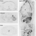
An adequate sequence design of diffusion-weighted sequence in the upper abdomen is critical, because it has intrinsically limited spatial resolution. Box 1 summarizes the list of scanning parameters to be optimized in a DWI sequence of the body. Most commonly, a single-shot spin-echo echo-planar imaging sequence is performed, which has the advantage of a very fast readout, making it insensitive to macroscopic patient motion. However, this family of sequences is prone to motion and magnetic susceptibility artifacts. The maintenance of echo time (TE) as short as possible, using parallel imaging, high bandwidth, and advanced suppression techniques, minimizes distortion artifacts, although diffusion requires intrinsically long TEs due to the time required to impart sufficient diffusion sensitivity. In order to reduce the effects of respiratory and cardiac motion, it is necessary to use gated acquisitions ( Fig. 1 ). Most commonly, breath-hold or free-breathing sequences are used ( Box 2 ). In addition, fat suppression must be used to avoid ghosting artifacts from the fat signal.
Increase SNR
Maintain TE as shortest as possible
Coarse matrix
Use parallel imaging
Simultaneous gradient application
Multiple signal averaging
Reduce artifacts
Optimize fat suppression
Increase bandwidth
Control eddy currents
Avoid areas with susceptibility artifacts
Use respiratory synchronism techniques
Breath-hold sequence
Limited SNR, spatial resolution, and slice thickness
Prone to distortion and ghosting artifacts
Limited number of b values (usually 2 or 3)
Limited quality of ADC map due to misregistration
Shorter acquisition time
Free breathing sequence
Increased SNR, spatial resolution, and slice thickness
Prone to respiratory artifacts, blurring, and volume averaging
Permit to acquire more b values
Better quality of ADC map
Usually longer acquisition time
Modeling of diffusion signal
In order to use DWI as an oncological biomarker, quantitative mapping is critical, especially in the setting of therapy monitoring. The most widely used quantitative property is the ADC from a monocompartmental model of diffusion signal decay. ADC measurement minimizes the so-called T2 shine-through, referring to the high signal from long T2 species seen in DWI because of superimposed T2 weighting and permits evaluation of isolated diffusion effects.
However, the ADC model does not distinguish between the different compartments where the water protons can move, such as intravascular, extravascular, extracellular, and intracellular spaces. If several b values are acquired less than and greater than 100 s/mm 2 , it is possible to differentiate between the fast movement of intravascular water molecules with low b values (<100 s/mm 2 ), and the slow signal decay of diffusion signal with b values greater than 100 s/mm 2 , secondary to restricted water movement in the extracellular and intracellular compartments (see Fig. 1 in the article by Broncano in this issue). This model is known as Intra Voxel Incoherent Motion (IVIM) because it has been found useful in the characterization of focal liver and pancreatic lesions, with advantage over ADC measurements in some scenarios. The contribution of true diffusion and perfusion toward signal loss is separated in this model and reflected in the following parameters: f (perfusion fraction) that represents the flowing water molecules within the capillaries; D (tissue diffusivity), a more reliable marker of tissue diffusion than ADC in organs with tissues with significant perfusion fraction; and finally, D* (pseudodiffusion coefficient), related to the perfusion contribution to signal decay.
If ultrahigh b values greater than 1500 s/mm 2 are acquired, the remaining diffusion signal is related to a layer of polarized water molecules near of the charges of the membranes. The measurement of this very slow diffusion pool requires the use of a non-Gaussian model, such as diffusional kurtosis imaging (DKI), which reflects the microstructural complexity of tissue (see Fig. 1 in the article by Broncano in this issue). Derived parameters from DKI can help in the characterization of focal liver lesions. Compared with ADC, these models provide supplementary information of the diffusion signal from other compartments different to extracellular one ( Fig. 2 ).
b-values selection
ADC calculation minimally requires the use of 2 b values, although the more b values obtained, the better the reliability of ADC maps. In the abdomen, typically the ideal higher b value ranges between 600 and 1000 s/mm 2 in order to maintain a sufficient signal-to-noise ratio (SNR). DKI requires a maximum b value in the range of 1500-2000 s/mm 2 , and IVIM analysis requires several b values less than and greater than 100 s/mm 2 , although it is feasible even with only 3 to 4 b values. However, the optimum set of b values and diffusion model used for analysis for oncological applications in the body is still to be defined.
Clinical Applications of Diffusion-Weighted Imaging
Liver
Lesion detection
DWI using a low b value around 50 s/mm 2 , also known as black-blood diffusion, allows better detection of small focal liver lesions against a liver parenchyma without vessel signal ( Fig. 3 ). This type of approach has been shown to improve detection of metastases in comparison to fat-suppressed T2-weighted sequences and is of special interest for identifying metastases from colorectal cancer before surgery, with particular advantage in the detection of lesions smaller than 1 cm and those adjacent to vascular structures. Furthermore, in patients at risk for nephrogenic systemic fibrosis, DWI can be considered an alternative to contrast-enhanced imaging. DWI in combination with gadoxetic acid (Gd-EOB-DTPA) improves detection of liver metastases compared with any individual technique alone, even after neoadjuvant chemotherapy.
Lesion characterization
The role of DWI in focal liver lesion characterization is controversial due to misregistration between images with different b values, caused by respiratory motion. Adequate reproducibility has been achieved for ADC, but lower for f and D∗. DWI has been shown to accurate differentiate between cystic and solid liver lesions. Benign lesions tend to show higher ADC values and lower SI with high b values than malignant ones ( Fig. 4 ). However, there are no definitive data to support the use of ADC in the distinction between benign and malignant focal liver lesions, due to substantial overlap, particularly focal nodular hyperplasia (FNH) and adenomas with hepatocellular carcinoma (HCC) and metastasis. In addition, radiologists must be alert to potential pitfalls such as low ADC values seen in pyogenic abscess or high ADC values of necrotic or mucinous metastasis.
IVIM-derived D and ADC show inconclusive results in the distinction of benign and malignant lesions, although with contradictory data about which of both parameters is more accurate. D* and f are useful to differentiate hypervascular from hypovascular lesions.
Hepatocellullar nodules in cirrhosis
DWI is of limited value in the evaluation of focal lesions in cirrhotic livers. DWI shows a lower sensitivity compared with dynamic contrast-enhanced MR imaging (DCE-MR imaging) and hepatobiliary (HB) phase in HCC detection. It also does not allow differentiation between dysplastic nodules and early HCC. However, DWI should be included in the protocol of detection of HCC, because it significantly improves sensitivity when used in combination with either technique. Most commonly, HCC shows restriction of water diffusion, paralleling degree of cellularity and dedifferentiation. Furthermore, a recent meta-analysis concluded that DWI had excellent and moderately high diagnostic accuracy for the prediction of well-differentiated versus poorly differentiated HCC, although overlap remains between different histologic grades. Recently, IVIM-derived D values of HCC showed significantly better diagnostic performance than ADC values in differentiating high-grade from low-grade HCC. In addition, f demonstrated a significant correlation with the percentage of arterial enhancement of HCC. Interestingly, benign lesions associated with cirrhosis, such as confluent fibrosis or perfusion alterations, do not show increased signal with high b values.
Therapy monitoring
DWI has been used for early assessment of tumor response to treatment of both primary and secondary malignancies of the liver ( Figs. 5 and 6 ). The good reproducibility of ADC measurement means that any ADC changes after treatment can be related to treatment effects. In general, increases in ADC after 1 week of successful treatment can be detected in the liver.
For example, DWI with ADC is useful in the early assessment of HCC response after transcatheter arterial chemoembolization (TACE), and differentiating between viable and necrotic portions of the treated HCCs. Furthermore, DWI can detect recurrent tumor after treatment and predict HCC response to TACE using the monoexponential or biexponential models of analysis.
DWI also can be used as a biomarker of treatment response of HCC to the antiangiogenic agent, sorafenib. First, ADC decreases probably related to hemorrhagic necrosis and, posteriorly, increases because of tumor necrosis. A delayed ADC decrease suggests tumor recurrence. IVIM-related D and f have been shown also to be valuable markers of treatment response to sorafenib for HCC.
For therapy monitoring of hepatic metastases, increases of ADC after the start of chemotherapy or radiotherapy have been related to responding metastases, although there is no defined threshold of increase in ADC to define response. These changes can be detected during the first week after the start of treatment in colorectal and breast liver metastases and occur before changes in size. In addition, DWI appears superior to positron emission tomography/computed tomography (PET/CT) for early response assessment of metastases of common solid tumors treated with Y90 radioembolization. Lower pretreatment ADC value has been related to good response to chemotherapy in colorectal and gastric hepatic metastases, although these data were not confirmed in a prospective series including digestive tract and breast liver metastases. Furthermore, low-pretreatment ADC value of colorectal liver metastases has shown an association to shorter overall survival and progression-free survival.
Biliary system and gallbladder malignancies
DWI detects intrahepatic and extrahepatic cholangiocarcinoma (CHC) with advantage over T2-weighted and in a similar manner to DCE-MR imaging. Because this lesion shows the lowest ADC of all hepatic malignancies, its differentiation from benign focal liver lesions using DWI is feasible. DWI also helps in the differentiation between mucus and viable intraductal CHC obstructing the bile duct. The lower the degree of tumor differentiation in CHC, the lower the ADC value is. A target appearance on DWI, a hyperintense peripheral halo (viable tumor) with a hypointense central area (fibrosis), permits the accurate differentiation of small intrahepatic CHC from HCC, because this sign is superior to other morphologic, DCE-MR imaging, or HB phase features.
As with other malignancies, gallbladder carcinoma typically demonstrates high signal on high b-value imaging, and low ADC values ( Fig. 7 ). Acute cholecystitis may simulate a neoplastic process on DWI. For this reason, DWI must be evaluated along with the other morphologic images, increasing the accuracy in the differentiation of benign and malignant gallbladder lesions.
Pancreatic cancer and other pancreatic tumors
A recent meta-analysis demonstrates high performance of DWI in detection of pancreatic adenocarcinoma (PA), with similar sensitivity to fluorodeoxyglucose (18FDG) PET/CT, but better specificity. Most commonly, PA shows high SI on high b-value imaging and lower ADC values than normal pancreas, related to dense fibrosis and increased cellular elements ( Fig. 8 ). However, PA can have different appearances on diffusion imaging depending on histologic characteristics. Edematous fibrosis and loose collagen fibers have been described in PA to yield higher ADC than normal parenchyma. This heterogeneity in histologic content is probably the cause of contradictory data in the relationship between ADC values of PA and tumor grade. Moreover, the association of acute pancreatitis and PA located in the pancreatic head has been reported. Both entities show a similar behavior in DWI, hampering cancer detection. The use of IVIM-derived parameters, such as f, improves the differentiation between PA and normal pancreatic tissue.
DWI is also helpful in the differentiation between PA and benign lesions, although with limitations in the distinction from mass-forming pancreatitis. There is an evident overlap in ADC values of both lesions, probably due to variable proportions of fibrosis and inflammation in mass-forming pancreatitis and different degrees of PA differentiation (see Fig. 8 ; Fig. 9 ). However, initial data suggest a role for IVIM-derived perfusion parameters in this distinction.
In the therapy monitoring setting, preliminary data suggest that lower pretreatment ADC values of advanced PA, treated with chemotherapy or chemoradiation, are related to poor response and early progression ( Fig. 10 ). DWI is also useful in the evaluation of autoimmune pancreatitis response to steroids. This entity shows high SI on high b value and lower ADC values than chronic pancreatitis and PA. However, with successful treatment, SI on high b value decreases, and ADC returns to normal values.
Pancreatic neuroendocrine tumors (PNET) show a restrictive pattern in diffusion, in comparison with normal pancreatic parenchyma ( Fig. 11 ). However, the detection rate of PNETs with MR imaging including DWI was significantly lower than that of 68 Ga-DOTATATE PET/CT. Most aggressive PNETs show lower ADC values than benign lesions. In addition, an inverse correlation between Ki-67 (cellular marker for proliferation) and ADC values has been established, which would allow for the evaluation of tumor aggressiveness. Perfusion parameters from IVIM models are able to accurately differentiate PNET from PA.
Pancreatic cystic lesions are a common incidental finding. It is important to differentiate cystic tumors from nonneoplastic lesions and identify malignant variants. Cystic tumors show different biological behavior and risk of malignancy and can be classified as shown in Box 3 . Cystic lesions greater than 2 cm in size are usually premalignant or malignant.
Benign cystic tumors
Serous cystadenoma
Cystic tumors with malignant potential
Mucinous cystic neoplasms (MCN)
Intraductal papillary mucinous neoplasm (IPMN)
Frankly malignant cystic tumors
Cystadenocarcinoma
Intraductal papillary mucinous adenocarcinoma (Malignant IPMN)
Cystic-appearing pancreatic tumors
Solid pseudopapillary neoplasm
Acinar cell cystadenocarcinoma
Lymphangioma
Hemangioma
Paraganglioma
Solid pancreatic lesions with cystic degeneration
Pancreatic adenocarcinoma
Cystic pancreatic neuroendocrine neoplasm (PNET)
Metastasis
Cystic teratoma
Sarcoma
Nonneoplastic cystic lesions
True epithelial cyst
Mucinous nonneoplastic cyst
Pseudocyst
Abscess
DWI with high b value can differentiate nonneoplastic cysts, such as simple cysts and pseudocysts, which are usually isointense to the pancreas, from neoplastic cysts and abscesses, which remain hyperintense. Boraschi and colleagues found statistically significant differences in ADC values of intraductal papillary mucinous neoplasms (IPMNs), mucinous cystic neoplasms (MCNs), serous cystadenomas, and pseudocysts, although other series have shown significant overlap in the ADC values of cystic pancreatic lesions. More consistent data have shown significantly higher ADC values for IPMNs compared with MCNs. Furthermore, ADC and D values of malignant IPMN are significantly lower than benign variants ( Figs. 12 and 13 ). Currently, the differentiation of cystic pancreatic lesions only based on DWI remains limited, although integrated in a comprehensive MR protocol, diffusion is considered helpful.
Perfusion MR imaging
A mainstay of tissue and lesion characterization in abdominal MR imaging is a DCE series, which assesses the enhancement of tissues at predetermined time points after contrast injection. The core physiology probed by this examination is perfusion, which is defined as the passage of blood through the capillary bed to deliver nutrients and oxygen to the tissue. The current clinical assessment uses 2 to 4 time points after contrast injection to visually assess enhancement patterns in organ parenchyma and lesions, which indirectly provide valuable information about tissue physiology and pathology. One limitation to this approach is that very few images are acquired to analyze a very complex enhancement curve. The arterial phase of enhancement, in particular, can be difficult to capture because of the transient and temporally variable nature of enhancement of lesions during this phase. Moreover, this assessment is qualitative, and factors, such as the radiologist’s skill, accuracy of timing after injection, and the protocol used, can inordinately affect the clinical reading. A major frontier in abdominal MR imaging is the development of an accurate quantitative DCE-MR imaging examination that enables extraction of perfusion parameters, which reflect tissue properties. Recent literature suggests that this quantitative approach has potential applications in early diagnosis of liver cirrhosis, noninvasive diagnosis of focal lesions, assessment of response to novel antiangiogenic chemotherapeutic drugs, and in predicting treatment response in tumors as HCC and PA.
Technical Design of Perfusion MR Imaging of Upper Abdomen
Image acquisition
The characteristics of an ideal DCE-MR imaging perfusion examination are listed in Box 4 . Gadolinium-based contrast agents (GBCAs) are used as tracers and image acquisition aims to accurately capture changes in tissue SI, which is related to changes in concentration of the GBCA over time. Both extracellular and hepatobiliary contrast agents (HBCA; Gd-EOB-DTPA) have been used for perfusion MR imaging studies. Before injecting the contrast agent, a T 1 quantification acquisition is performed to determine the baseline tissue signal. This T 1 quantification acquisition is done by using the variable flip angle technique ; flip angles in the range of 30° to 60° are recommended. This T 1 quantification acquisition is followed by the actual DCE-MR imaging acquisition: a GBCA (0.1 mmol/kg body weight) is injected through a wide-bore cannula (20 G or larger) placed in a large antecubital vein using a power injector at a flow rate of 2 to 4 mL/s, followed by a 20 to 30 mL saline flush. Repeated T 1 -weighted gradient echo imaging of the tissue of interest is performed every few seconds. The capillary transit time in well-perfused organs such as the liver is on the order of 3 to 5 seconds; hence, a sampling interval of less than 2 seconds per volume is typically required. Multiple volumes are acquired over a period of 3 to 5 minutes, starting 10 to 20 seconds before contrast injection, to capture the change in concentration of the GBCA with time.
Related posts:
 Body Diffusion-weighted MR Imaging in Oncology
 Assessment of Tumor Angiogenesis
Clinical Imaging of Tumor Metabolism with 1H Magnetic Resonance Spectroscopy
Multiparametric MR Imaging of Breast Cancer
Role of Multiparametric MR Imaging in Malignancies of the Urogenital Tract
Functional MR Imaging in Gynecologic Cancer
Assessment of Tumor Angiogenesis
Clinical Imaging of Tumor Metabolism with 1H Magnetic Resonance Spectroscopy
Multiparametric MR Imaging of Breast Cancer
Role of Multiparametric MR Imaging in Malignancies of the Urogenital Tract
Functional MR Imaging in Gynecologic Cancer
Stay updated, free articles. Join our Telegram channel

Full access? Get Clinical Tree


