, Valery Kornienko2 and Igor Pronin2
(1)
N.N. Blockhin Russian Cancer Research Center, Moscow, Russia
(2)
N.N. Burdenko National Scientific and Practical Center for Neurosurgery, Moscow, Russia
Affecting the central nervous system, a fungal infection is regarded as an opportunistic granulomatous disease that can be acute and fulminant or chronic and slow. Fungi can invade the central nervous system and cause meningitis or parenchymal focal lesions in some patients suffering from systemic fungal infections.
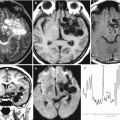
MRI and CT manifestations of fungal infections are similar to those of tuberculosis (Lyons and Andriole 1986; Sze et al. 1998). CT and MRI can identify a space-occupying brain lesion, abscesses, infection foci in the eye sockets or sinuses, and hydrocephalus. The most common fungi are divided into opportunistic microorganisms that affect only immunocompromised patients (Candida, Aspergillus, Mucor) and microorganisms that also infect immunocompetent individuals. Cryptococcosis, histoplasmosis, and blastomycosis may arise both in previously healthy individuals and in patients with compromised immunity. Candida, Aspergillus, Mucor, and Cryptococcus neoformans live worldwide; the rest are endemic to certain geographical areas of the world. In addition, pathophysiology of fungal infections of the central nervous system depends on the fungus species. Fungi that reproduce as yeast (by division) enter the CNS via the hematogenous route (Cryptococcus and Histoplasma), enter the microvascular system of meninges, and penetrate the vascular wall, causing acute or chronic meningitis. Intracerebral lesions, such as granulomas or abscesses, are less common (Gaviani et al. 2005).
Fungi that reproduce in the infected tissue by hyphae (Aspergillus, Mucor) or pseudohyphae (Candida) tend to invade the parenchyma, not the meninges, since the blood supply of meninges is insufficient for them. Hyphae that form fungal colonies are able to penetrate and occlude large, medium, and small arteries, resulting in cerebral vascular accidents and cerebritis (Table 43.1).
Table 43.1
Pathogens of fungal meningitis
Pathogen | Opportunistic | Other affected structures | Characteristic changes in CSF |
|---|---|---|---|
Cryptococcus neoformans | Sometimes in AIDS | Lungs, bones, joints | Viscous consistency, stained with ink, positive reaction to anti-cryptococcal antigen |
Coccidioides immitis | No | Lungs, skin, bones | Positive reaction of complement binding |
Candida | Yes | Mucous membranes, skin, esophagus, urinary tract, heart
Related posts:Stay updated, free articles. Join our Telegram channel
Full access? Get Clinical Tree
 Get Clinical Tree app for offline access

|