One application that requires a slow compression is poroelastography, in which the flow of interstitial fluid under compression, which occurs over several seconds, is measured. The clinical analogy is the demonstration of pitting edema, for example, over the ankle, shown by sustained gentle finger compression and then palpation of the resulting pitting of the skin. The problem has been modeled mathematically.1,2 An ultrasound application has been described in transplant kidneys, where steady probe compression and relaxation was applied in 3 second cycles and speckle tracking was used to assess the resulting distortion.3 Significant differences were obtained in the relaxation times compared to the Banff scores indicating differences in edema.
New ways to displace the tissues under examination include sending several acoustic radiation force impulses (ARFIs) from different parts of the array simultaneously. Known as comb push elastography it allows higher frame rates than achievable with a single push pulse. The overlap of several shear waves leads to interference patterns that may reveal new tissue properties.4
The shear waves produced by conventional ARFI push pulses are currently assumed to travel at a single speed, which is used to assess tissue stiffness. But this is a simplification. Actually shear waves occupy a range of frequencies from around 50 to 500 Hz. Those of higher frequencies travel faster than those of lower frequencies (unlike compressional audible sound and ultrasound waves, in which all frequencies travel at the same speed). This phenomenon, known as dispersion, seems to result from properties of the viscous component of the tissue. Understanding how each manufacturer’s shear wave system deals with this dispersion could lead to improvements in the spread of reading due to improved consistency across systems.5 Estimation of the dispersion has been shown in ex vivo animal and human studies to allow diagnosis of the amount of fat in the liver and could provide information on the degree of steatosis, which is a major clinical problem.6
The concept of harnessing the shear waves that are generated throughout our bodies by native cardiovascular movements is the subject of active research.7 The challenge is to detect these low amplitude shear waves from noise and to decipher the complex interference patterns produced when they overlap. It could open the way to a wider range of shear wave speed estimations.
13.3 Optical Methods
Optical coherence tomography (OCT) is powerful technique for imaging superficial structures such as the skin and the eye using laser light at visible wavelengths and a phase-sensitive technique to produce tomograms with submicron resolution. It has become a standard technique for examining the retina and subretinal layers in ophthalmology and can be adapted as a strain elastographic method by comparing OCT scans taken before and after a small distorting force is applied; for example to the cornea by a puff of air (as used to measure intraocular pressure in glaucoma).8 An important potential application is in keratoconus, in which the cornea softens and projects anteriorly. Sufferers experience progressive changes to their refraction, and may be offered laser treatment, which has the disastrous effect of further weakening the cornea.9
Optical coherence elastography has been developed to assess the mechanical properties of the skin in tumors and in burns.10,11 Using a probe with a transparent membrane to contact the skin and apply a distorting force (which could be manual, in a handheld device, or automatic, using a piezoelectric crystal), phantom and clinical studies have been performed. The use of a 1mm thick Elastosil transparent stress sensor plate between the probe and the skin to measure the applied stress allows quantitative estimates of the skin’s elasticity. The trial images have strikingly high strain contrast at submillimeter resolution. If similar stress sensors could be incorporated into ultrasound transducers, quantitative strain ultrasound elastography might become possible.
13.4 Optoacoustic Methods
When a coherent light beam traverses or is back-scattered from a material that is vibrated mechanically, the movement of the material disturbs the light beam and changes the laser speckle pattern. A camera looking at the laser detects a reduction in the laser speckle contrast. This effect can be harnessed to sense the arrival of a shear wave, forming an alternative detection system to ultrasound with the advantage of better sensitivity than ultrasound offers.
The approach has been developed into an experimental shear wave system in which one or more ARFI pulses are directed into a test object and the time-of-flight of the shear wave is measured optically with great accuracy.12 In the experimental system, the camera watches for the transient reduction in laser speckle contrast while an ARFI pulse is sent into the material some distance to one side; repositioning the ARFI pulse can be used to measure the shear wave speed. Both theoretical models and simulation have been developed for the system. Like all optical systems, it is depth limited and, even using the longer wavelengths of infrared light, only 20 to 30mm of penetration can be achieved, so clinical use would be limited to structures close to the probes such as small parts and endoscopic applications.
13.5 New Clinical Applications
There are several clinical applications that are being explored but are not yet in clinical use. The literature regarding the most promising of these are summarized below.
13.5.1 Obstetrics and Gynecology
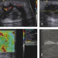
One of the most compelling possible clinical uses is assessment of the softening of the uterine cervix before delivery in late pregnancy (▶ Fig. 13.1). In the normal process, as well as in cervical shortening, there is a reduction in the cervix’s stiffness that allows cervical dilatation to permit passage of the fetal head along the birth canal. These changes are conventionally assessed by a combination of transvaginal ultrasound and manual palpation; these form the basis for the Bishop’s score that is used in obstetric decision-making. If this ripening occurs early, premature delivery is more likely, with devastating consequences for the neonate.
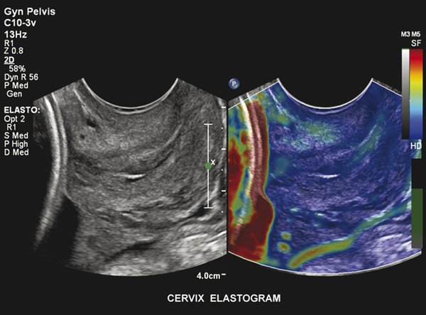
Fig. 13.1 Uterine cervix. Part of the stiff (red coding) fetal head can be discerned on the left of this strain elastogram taken with a transvaginal probe. The endocervix is slightly stiffer than the muscular portion, indicating that ripening has not progressed. (Courtesy of Dr. John MacQuarry, Philips Medical Systems, Bothel, Washington.)
Attempts to use strain elastography have not proved very successful, probably because of the complex structure of the cervix, with intertwined layers of fibrous tissue and smooth muscle, and the difficulty of applying the necessary force uniformly.13 The shear wave approach seems more promising, though this may necessitate the development of special small transducers that can be applied directly to the posterior surface of the cervix via the posterior fornix.14
Another obstetrical application that is promising is in ectopic gestations, where suspicious adnexal masses that proved to be ectopic showed increased stiffness on strain elastography (termed the blue eye sign).15 This was reliable regardless of the β-HCG (beta-human chorionic gonadotropin) level or the conventional ultrasound features.
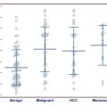
The stiffness of predominantly cystic ovarian masses has been studied in a small group of patients (26) using transvaginal strain elastography and a subjective scoring system.16 Most lesions showed the mixed pattern suggestive of simple cysts, but 3 of the masses contained stiffer regions and these corresponded to carcinomas, as confirmed on histology. The method could be a useful adjunct to conventional transvaginal assessment when the diagnosis of ovarian cancer is entertained.
Polycystic ovaries (PCO) were found to be stiffer than normal ovaries on strain elastography in a study of 48 patients compared with an equal number of healthy volunteers,16 and the interobserver correlations were excellent. Measurement of ovarian stiffness merits further investigation as a means to improve the diagnosis of PCO in conjunction with ovarian appearance and volume.
A pilot study of over 200 women (some normal and others with a variety of uterine pathologies) using transvaginal strain elastography demonstrated that the normal myometrium had a uniform, stiff appearance that was distinct from the serosa, which presented a laminar pattern.17 Leiomyomas (56 cases, confirmed on histology or MR) were also found to be uniformly stiff but somewhat stiffer than normal myometrium (▶ Fig. 13.2). Adenomyomatosis (11 cases, confirmed on MR) were softer than myometrium with an irregular pattern and margins. Some artifacts that are important to recognize were highlighted in the study, as was the need for practice in order to obtain repeatable results.
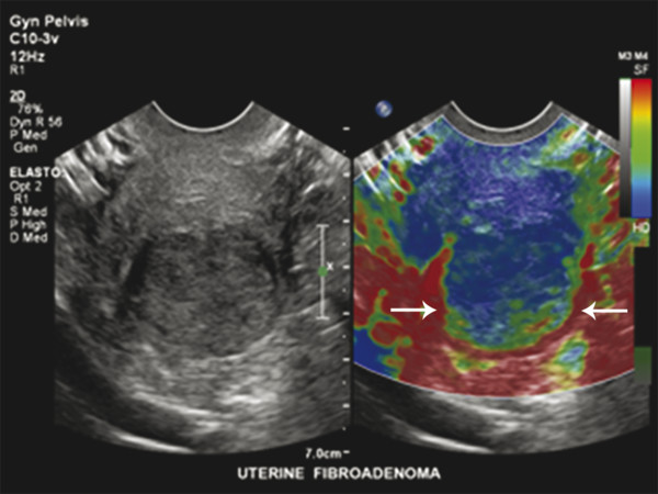
Fig. 13.2 Uterus with fibroid. In this transvaginal strain elastogram of the uterus, the myometrium has a uniform, rather stiff appearance while the fundal fibroid (arrowheads) is stiffer and somewhat heterogeneous. (Courtesy of Dr. John MacQuarry, Philips Medical Systems, Bothel, WA.)
In a case report of a women with a leiomyosarcoma compared with another with a simple leiomyoma (fibroid), the malignancy was found to be stiffer on strain and shear wave elastography, both performed using an ARFI method.18 The leiomyosarcoma also showed more heterogeneity of stiffness through the lesion. Larger studies are needed.
13.5.2 Spleen
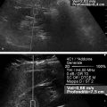
The spleen’s mechanical properties are affected by the hemodynamic changes that occur in portal hypertension, and this has been proposed as an alternative to assessing the liver itself, especially where the liver is difficult to evaluate because it is badly damaged and heterogeneous (▶ Fig. 13.3). In a study of 123 patients with varying degrees of liver fibrosis and cirrhosis (mainly attributable to hepatitis C), using the SuperSonic imaging (SSI) shear wave elastography system (Aixplorer, SuperSonic Imagine), spleen stiffness was higher than liver stiffness at all grades.19 A cutoff value between mild and severe liver fibrosis of 23 kPa (cf., 11 kPa for normal liver) is recommended. A high body mass index (BMI) and small spleen size were associated with failed spleen measurements, which were more common than failed liver measurements.
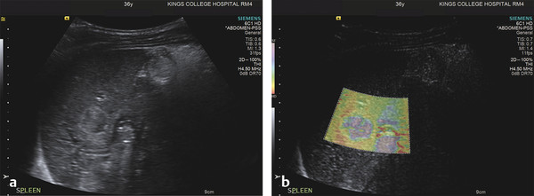
Fig. 13.3 Splenic abscess. A 36-year-old male recently arrived from the Middle East presented with fever and left upper quadrant pain. The B-mode scan (a) showed a well-defined, solid-appearing heterogeneous splenic mass. On ARFI strain elastography, it was found to be of mixed medium-to-soft stiffness (green-to-mauve) in comparison to the surrounding spleen, which was also somewhat heterogeneous. It proved to be a bacterial abscess, caught in the preliquefactive phase. (Courtesy of Prof. Paul Sidhu, King’s College Hospital, London, UK.)
Related posts:
Stay updated, free articles. Join our Telegram channel

Full access? Get Clinical Tree


