Fig. 7.1
A schematic diagram of a gas-filled detector illustrating the principles of operation
At very low voltages, the ion pairs do not receive enough acceleration to reach the electrodes and therefore may combine together to form the original molecule instead of being collected by the electrodes. This region is called the region of recombination (Fig. 7.2). As the applied voltage is gradually increased, a region of saturation is encountered, where the current measured remains almost the same over the range of applied voltages. In this region, only the primary ion pairs formed by the initial radiations are collected. Individual events cannot be detected; only the total current passing through the chamber is measured. Because specific ionization differs for α-, β-, and γ-radiations, the amount of current produced by these radiations differs in this region. The voltage in this region is of the order of 50–300 V. Ionization chambers such as dose calibrators are operated in this region.
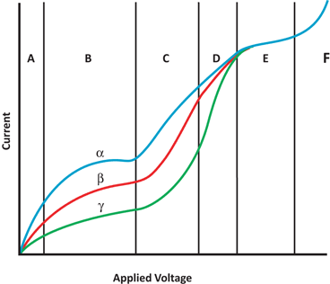
Fig. 7.2
A composite curve illustrating the current output as a result of increasing voltages for different radiations. a Region of recombination, b region of saturation, c proportional region, d region of limited proportionality, e Geiger region, and f continuous discharge
When the applied voltage is further increased, the electrons and positive ions gain such high velocities and energies during their acceleration toward the electrodes that they cause secondary ionization. The latter will increase the measured current. This process is referred to as the gas amplification . This factor can be as high as 106 per individual primary event depending on the design of the gas detector and the applied voltage. In this region, the total current measured is equal to the number of ionizations caused by the primary radiation multiplied by the gas amplification factor. In this region, the current increases with the applied voltage in proportion to the initial number of ion pairs produced by the incident radiation. Therefore, as in the case of the region of saturation, the current amplification is relatively proportional to the types of radiations, e.g., α-, β-, and γ-radiations. This region is referred to as the proportional region (see Fig. 7.2). Proportional counters are usually filled with 90 % argon and 10 % methane (P-10 ) at atmospheric pressure. These counters can be used to count individual counts and to discriminate radiations of different energies, but are not commonly used for γ– and x-ray counting because of poor counting efficiency (<1 %).
As the applied voltage is increased further, the current produced by different types of radiation tends to become identical. The voltage range over which the current tends to converge is referred to as the region of limited proportionality . This region is not practically used for detecting any radiation in nuclear medicine.
With additional increase in voltage beyond the region of limited proportionality, the current becomes identical for all radiations, regardless of how many ion pairs are produced by the incident radiations. This region is referred to as the Geiger region (see Fig. 7.2). In the Geiger voltage region, the current is produced by an avalanche of interactions. When highly accelerated electrons strike the anode with a great force, ultraviolet (UV) light is emitted, which causes further emission of photoelectrons by gas ionization and from the chamber walls. The photoelectrons will again strike the anode to produce more UV, and hence an avalanche spreads along the entire length of the anode. The amplification factor can be as high as 1010. During the avalanche, however, the lightweight electrons are quickly attracted to the anode, whereas a sheath of slow-moving heavy positive ions builds up around the anode. As a result, the voltage gradient falls below the value necessary for ion multiplication, and therefore the avalanche is terminated. All this occurs in less than 0.5 µs, and the counter is left insensitive and must recover before another event can be counted.
Recovery begins with the migration of the positive ions toward the cathode (i.e., chamber wall) and takes about 200 µs at a gas pressure of 0.1 atmosphere, which is equal to the dead time of the counter that varies with gas pressure. As the positive ions approach the cathode, secondary electrons may be emitted from the surface of the cathode, which then set another discharge just about 200 µs after the previous one. Such repetitive discharges that are due to secondary electrons are independent of the types and energy of radiation that the counter is intended to measure. The emission of secondary electrons is suppressed by a technique known as quenching to eliminate repetitive counter discharges (see later).
As the applied voltage is increased beyond the Geiger region, a single ionizing event produces a series of repetitive discharges leading to what is called spontaneous discharge . This region is called the region of continuous discharge because the gas may be ionized in the absence of radiation at this high voltage (see Fig. 7.2). Operation of a detector in this region may cause damage to the detector.
Ionization Chambers
Ionization chambers are operated at voltages in the saturation region that spans 50–300 V. The detector is a cylindrical or rectangular chamber filled with air or a gas , sometimes at high pressure. A central wire and the chamber act as the electrodes and the current is measured by an electrometer. The detection efficiency of the ionization chambers for x-rays and γ-rays is very low (<1 %) and depends on the energy of these radiations . Ionization chambers are primarily used for measuring high-intensity radiation such as x-ray beams and high activity of radiopharmaceuticals. Ion chamber survey meters , dose calibrators , and pocket dosimeters are the common ionization chambers used in nuclear medicine.
Ion Chamber Survey Meter
An ion chamber survey meter consists of a metallic box fitted with a voltage circuitry operated by batteries to measure the ionization current or ion-pairs produced by interaction of radiations with the gas molecules in the chamber. The readings are displayed in analog or digital mode on scales over four or five decades (background to 50 R/h). They can be used to measure α, β and γ-ray exposure rates; however, for measurement of γ-ray exposure alone, a retractable beta shield (1000 mg/cm2) is placed beside a thin window (7 mg/cm2) of the chamber. The shield is removed when measuring α and β particle exposures. However, in the case of α particle exposure, the pulse mode counting is applied due to large voltage pulses produced, whereas in the case of β and γ rays, the current mode is employed. Most meters are operated at ambient atmospheric pressure, whereas in some, high pressure is employed using argon or similar gas. In the latter, the probability of photon interaction increases and hence the sensitivity. However, the operation of the meter at atmospheric pressure is affected by variations in temperature and atmospheric pressure at different geographical locations causing a change in gas density, and so readings will differ from the calibration value. Correction circuits are installed in current ion chambers to correct for the temperature and pressure changes. A typical ion chamber is shown in Fig. 7.3.
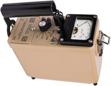
Fig. 7.3
An ion chamber survey meter (Courtesy of Ludlum Instruments, Inc. Sweetwater, TX)
According to NRC regulations, the ion chambers must be calibrated annually with a calibration source . 137Cs (t1/2 = 30 year) with 662 keV photons is commonly used for calibration of these ion chambers and other survey meters. The sensitivity of these meters is quite linear over a broad range of photon energies relative to 137Cs response except at low energies (Fig. 7.4). These meters can be used for a wide range of exposure readings except at low energies. The output is integrated and averaged over a period of time by the use of an RC circuit, and integration can be made slower or faster by the use of a switch.
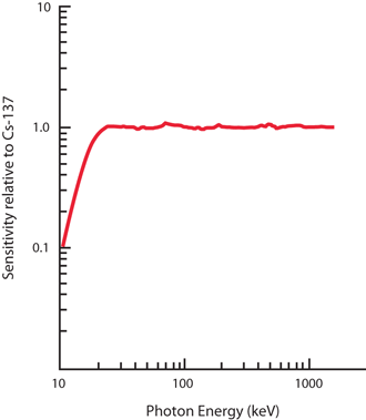
Fig. 7.4
The relative energy-independent response of ionization chamber
Standard ion chambers are used for measurement of moderate to high exposure rate readings (e.g., 99Mo–99mTc generator, X-ray beam intensity etc.) , whereas pressurized ion chambers can be used for measuring low exposure rate readings. Sometimes scintillation detectors that are fabricated in the form of a probe are also used to measure the exposure rates in low to moderate range.
Related posts:
Stay updated, free articles. Join our Telegram channel

Full access? Get Clinical Tree


