Near gastroesophageal (GE) junction, on posterior aspect of lesser curvature of stomach


 with an air-contrast level seen within an outpouching near the gastric cardia.
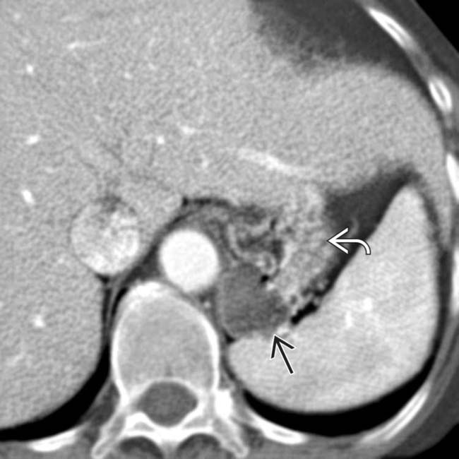
 projecting posterior to the gastric fundus
  . The connection to the stomach is much more difficult to see on CT. Distention of the stomach with oral contrast or gas granules may be required to make the diagnosis on a CT scan.
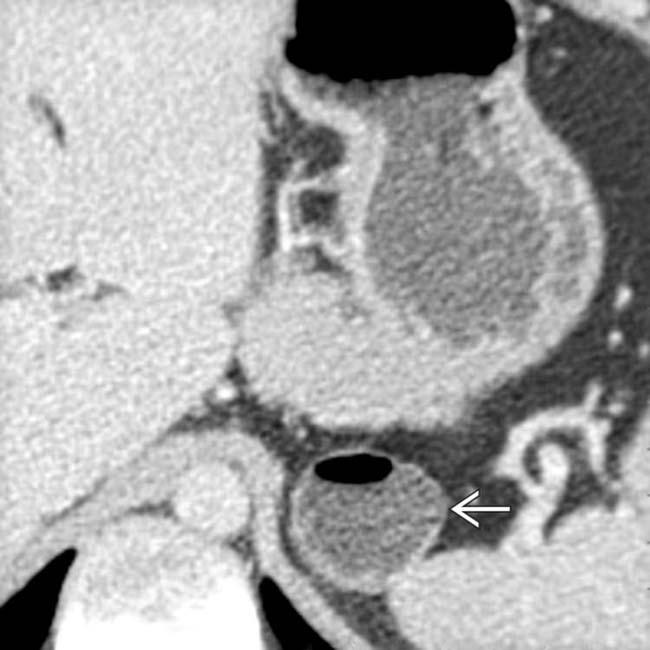
 containing water density fluid and gas. On more cephalic sections, the “mass” was contiguous with the posterior wall of the fundus.
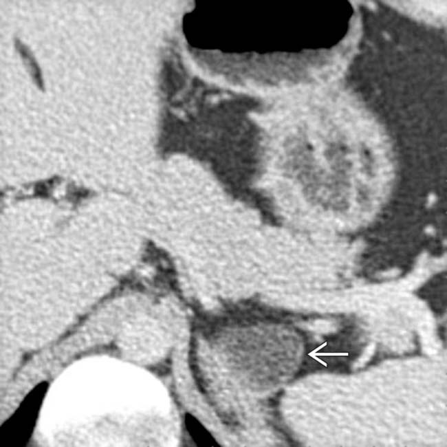
 extends dorsal to the pancreas and splenic vein. Without the presence of the air-fluid level it would be difficult to distinguish this from an adrenal mass. An upper GI series confirmed a typical juxtacardiac diverticulum.
IMAGING
General Features
• Best diagnostic clue


Related posts:
Stay updated, free articles. Join our Telegram channel
Full access? Get Clinical Tree
