84 Geometric Distortion
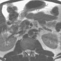
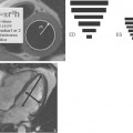
Figure 84.1A and Fig. 84.2A have substantial image distortion, the first at the bottom of the image and the second at the top. This results in the femur being artificially curved medially (arrow) in Fig. 84.1A and the vertebrae of the upper lumbar spine appearing progressively smaller (arrow) in Fig. 84.2A. In each case, the image distortion was due to nonlinear gradients. Virtually all MR systems suffer from some gradient nonlinearity, oftentimes due to coil design that seeks to optimize other aspects of gradient performance. It may be difficult on any one MR system to actually obtain an image depicting such spatial distortion, because postprocessing techniques may be employed unbeknownst to the user to correct the appearance of the image (Fig. 84.1B and Fig. 84.2B).
Stay updated, free articles. Join our Telegram channel

Full access? Get Clinical Tree


