100 Gradient Moment Nulling
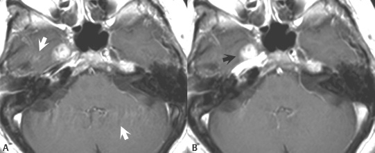
Fig. 100.1
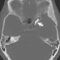
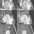
Figure 100.1A and Fig. 100.2A present spin echo contrast-enhanced T1-weighted and noncontrast T2-weighted images, respectively, with prominent motion artifacts (white arrows) in the phase-encoding direction. With the use of gradient moment nulling (GMN), artifact is markedly reduced and signal intensity is returned to dynamic structures of interest such as blood vessels (Fig. 100.1B) and cerebrospinal fluid (CSF) (Fig. 100.2B). The enhancing lesion (black arrow) in Fig. 100.1B, a benign nerve tumor (trigeminal schwannoma), is substantially more apparent on the image with GMN, due to reduced ghosting from the carotid arteries and transverse sinuses. Note that the use of GMN markedly improves depiction of CSF in Fig. 100.2B, which is now seen with uniform high signal intensity both anterior and posterior to the cervical cord.
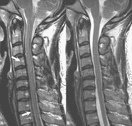
Fig. 100.2
Stay updated, free articles. Join our Telegram channel

Full access? Get Clinical Tree


