Figure 11.1
Endocavitary transducer. Endocavitary transducer, also known as transvaginal transducer

Figure 11.2
Transducer position for sagittal images. To obtain sagittal views of the uterus, the “trigger” or transducer marker should be pointed toward the floor

Figure 11.3
Transducer position for transverse images. To obtain transverse views of the uterus and ovaries, the “trigger” or transducer marker is pointed toward the patient’s left

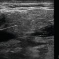
Figure 11.4
Bladder in sagittal view. In a sagittal plane using an endovaginal approach, the bladder (star) will be seen anterior to the uterus or on the left of the image. The uterine stripe is seen traversing the center of the uterus

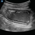
Figure 11.5
Long axis of uterus. The uterus is imaged in a sagittal plane from cervix (on the right of the image) to fundus (left of the image) with the echogenic uterine stripe in the midline

Figure 11.6
Pouch of Douglas. Posterior to the uterus and anterior to the rectum is the posterior cul-de-sac or pouch of Douglas. A small amount of free fluid is visualized here within the pouch of Douglas (star), which can be normal in reproductive-aged females

Figure 11.7
Uterus in transverse. In a transverse plane, the uterus is seen in short axis with the echogenic uterine stripe visualized as a small horizontal line within the center of the uterus

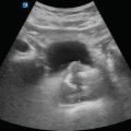
Figure 11.8
Normal ovary. Multiple small cysts (arrows) in the cortex surround the medulla (star) in this normal ovary, giving the appearance of what is often referred to as a “chocolate chip cookie”
Gynecologic Pathology
Simple Cyst
A simple ovarian cyst is a collection of fluid surrounded by a membrane within or on the surface of an ovary.
Simple cysts are anechoic with smooth walls, posterior acoustic enhancement, and no internal echoes:
Referred to as a physiologic cyst when the diameter measures less than 2.5 cm and a follicular cyst when it is greater than 2.5 cm [1]
Figure 11.9—Simple physiologic cyst

Figure 11.9
Simple physiologic ovarian cyst. When the diameter of a simple ovarian cyst measures less than 2.5 cm, it is referred to as a physiologic cyst
Figure 11.10—Simple follicular ovarian cyst

Figure 11.10
Simple ovarian cyst. When the diameter of a simple ovarian cyst measures greater than 2.5 cm, it is referred to as a simple follicular cyst
Video 11.5—Simple ovarian cysts
Polycystic Ovarian Syndrome
Syndrome that occurs due to imbalance of reproductive hormones with an unknown cause
Will appear as multiple small, usually less than one centimeter, simple cysts along the periphery of the ovary:
Complex Cyst
Get Clinical Tree app for offline access
A complex ovarian cyst contains both fluid and solid components:
Figure 11.12—Complex ovarian cyst.