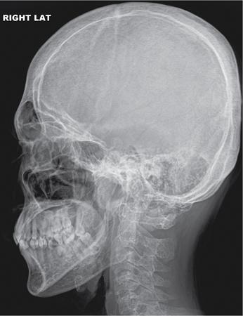
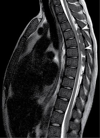
K.V. Rajagopal Haemoglobinopathy relates to the inherited disorders, which result in abnormal structure of one of the globin chains of the haemoglobin molecule. In these disorders, the RBCs are rounded instead of biconcave morphology and thus prone to destruction. Increased RBCs destruction would result in increased production of RBCs from bone marrow, which results in marrow hyperplasia. In later stages, it may result in extramedullary haemopoiesis. The common haemoglobinopathies encountered are thalassaemia and sickle cell anaemia. The radiographic and imaging features in haemoglobinopathies are mainly related to the marrow hyperplasia and extramedullary haematopoiesis. Few radiographic and imaging features are specific to certain haemoglobinopathy. Thalassaemia was first described by Cooley et al. (1927). Epidemiology: Thalassaemia is commonly encountered in Mediterranean countries, Southeast Asia, Northeast India. These conditions are generally inherited as an autosomal recessive pattern. Pathogenesis: Thalassaemia is classified into different types depending on the type of deficient chain involved: α, β, γ and δ. There is inadequate synthesis of beta-globin in β-thalassaemia, and there is inadequate synthesis of α-globin in α-thalassaemia, which would result in hypochromic microcytic anaemia. The ineffective production of RBCs results in osteoporosis, marrow expansion in the long bones, skull, ribs and facial bones, and it also leads to extramedullary haemopoiesis. The regular blood transfusions may lead to parenchymal iron deposition. Abnormal deposition of iron causes cirrhosis, cardiac dysfunction and endocrinopathies. Thalassaemias involving delta, gamma, epsilon and zeta chains are rare and are not associated with significant disease. Clinical features: Thalassaemia majorly results in severe childhood anaemia, hepatosplenomegaly, extramedullary erythropoiesis and skeletal deformity (e.g. rodent facies). Regular blood transfusions as required to maintain haemoglobin level, desferrioxamine (DFX) to treat iron overload and bisphosphonates prevent osteoporosis. Treatment complications related to transfusions and that related to DFX therapy must be familiar and diagnosed. The radiographic features in untreated thalassaemia major are secondary to marrow hyperplasia, osteopenia and extramedullary haemopoiesis. Medullary space all tubular bones are expanded with thin cortices, coarsening of the trabeculae with loss of normal tabulation, leading to squaring of the long bones. Extramedullary haematopoiesis denotes the development of erythropoietic tissue outside the bone marrow. Radiographic features are usually seen after the age of 1 year. The skull shows significant marrow expansion with a widening of the diploic space with thinning of the outer table (Fig. 2.4.1). The occipital bone is relatively spared. It may show a “hair-on-end” appearance due to the perpendicular orientation of the trabeculae. Paranasal sinuses are partially obliterated due to marrow hyperplasia. Spine also shows wide medullary spaces with coarsening of the trabeculae. Significant osteoporosis can lead to biconcave vertebrae and fractures of vertebral bodies. There can be associated with paravertebral soft-tissue masses and nodular extradural soft-tissue lesions due to extramedullary hemopoiesis. Extramedullary haemopoiesis in the extradural space can cause cord compression, which is best detected on MRI. Besides, MRI shows diffuse red marrow signal intensity in the vertebral bodies (Fig. 2.4.2). Chest X-ray shows posterior rib expansion, osteopaenia and adjacent soft-tissue thickening due to extramedullary haemopoiesis (Fig. 2.4.3).
2.4: Haemoglobinopathy
Thalassemia
Radiographic features


Stay updated, free articles. Join our Telegram channel

Full access? Get Clinical Tree


