Heart and Coronary Arteries
The heart is an organ that is asymmetrically situated in the mediastinum. It is protected by a mesodermal derived structure, the pericardial sac, consisting of two sheets: the external or parietal, which is fibrous, and the internal or visceral, which is a thin serous membrane attached to the heart muscle surface.
The heart is located in the middle portion of the inferior mediastinum bordered laterally by the medial face of the lungs, anteriorly by the chest wall, and posteriorly by the dorsal spine. In the majority of individuals, a large part of the heart lies in the left hemithorax and is partially covered by the lingula of the left lung (Figs. 13.1, 13.2, 13.3).
In humans, the heart is a double valvular pump that works physiologically in serial sequence. The superior and the inferior venae cavae drain the systemic venous blood into the right atrium, which is connected to the right ventricle through the tricuspid valve. The right ventricle pumps the blood to the pulmonary artery across the pulmonary valve. The arterial blood returning from the lungs drains into the left atrium through two left and two right pulmonary veins. The atrioventricular ring, where the mitral valve is attached, separates the left atrium from the left ventricle, which ejects the blood into the aorta across the aortic valve (Fig. 13.4).
The longitudinal axis of the heart is most frequently oriented anteriorly, inferiorly, and toward the left. If oriented toward the right, it characterizes the so-called dextrocardia, and if the longitudinal axis is in the sagittal plane and the heart occupies the middle mediastinum, it is called mesocardia (Fig. 13.5).
When the heart is faced in frontal view, the right border is formed by the lateral wall of the right atrium. The left margin is outlined cranially by the left atrium appendage and caudally by the lateral wall of the left ventricle (Figs. 13.1, 13.2, 13.6A). The right atrium and the right ventricle are ventral to the left atrium and the left ventricle is dorsal (Figs. 13.1, 13.6B, 13.7).
The heart has roughly the form of an inverted pyramid, with the base, formed by right and left atria and the root of the great vessels, located at the upper mediastinum. The apex of the pyramid corresponds to the apex of the left ventricle and is situated in the left hemithorax toward the diaphragm. The anterior face of the heart or sternocostal face is related to the anterior wall of the chest. The inferior face lies on the diaphragm and the lateral or pulmonary face is covered by the lingula of the left lung (Figs. 13.5, 13.8). The junction of the sternocostal face with the inferior or diaphragmatic face defines a sharp border called the acute margin. On the left side, the junction of the left face with the diaphragmatic face is the obtuse margin with a rounded and ill-defined border (Fig. 13.9). The divisions of the four cardiac chambers are seen externally as sulci or grooves. Between the atria and ventricles, there is the atrioventricular sulcus divided in right and left. The ventricle’s separation is marked externally by the interventricular sulcus, which is divided into anterior and inferior. The point at which the atrioventricular sulcus meets the inferior interventricular sulcus is termed the crux of the heart.
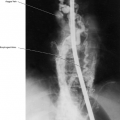
Although anatomic dissection has been the historical method for defining and advancing our understanding of human anatomy, medical imaging now plays an invaluable role. In the latter half of the twentieth century, angiography was the primary means of visualizing the cardiac structures in a living person. Although this was and is an excellent technique, it has two limitations. First, it is an invasive procedure requiring catheterization of the blood vessels with placement of a catheter into the heart and coronary arteries. Second, only the column of contrast material shows up on the radiographs, so it is the lumen of the arteries and cardiac chambers that is imaged. Angiography gives no direct information about the solid cardiac structures such as myocardium, pericardium, and valves. More recently echocardiography, magnetic resonance imaging, and multislice computerized tomography have become advanced to the degree that they provide a noninvasive means of evaluating cardiac anatomy. In addition to being noninvasive, these techniques provide additional information about all cardiac structures. Computerized tomography may ultimately prove to be the most useful, since it also visualizes the pulmonary structures very well.
Cardiac Chambers
In angiography, the axial projections for identification of the cardiac structures are used. These projections permit the visualization of the septum and the surrounding structures of the heart in more detail than the classical frontal and lateral views.
The routine of the angiographic study of the heart consists of three projections:
Long axial view—30° craniocaudal image intensifier inclination and 60° left anterior oblique patient inclination
Elongated right anterior oblique view—30° craniocaudal inclination of the image intensifier and 30° right anterior oblique patient inclination
Four-chamber view—30° craniocaudal image intensifier inclination and 30° left anterior oblique patient inclination
Additional views may be necessary in special situations. Frontal and lateral views may be the elected projections for adequate visualization of the interventricular septum in some complex cardiac defects. The pulmonary trunk and its bifurcation are better visualized in the sitting up projection with the patient lying supine and the image intensifier rotating 30° cranially.
In CT, the initial images presented are in an axial plane. Subsequently this information can be reformatted into any plane that is desired. Coronal and sagittal images are usually reconstructed along with the long and short axis views that have been used historically in echocardiography. In particular, the short axis left anterior oblique (LAO) view can be used to evaluate the left ventricle through the cardiac cycle, thereby allowing for computation of an ejection fraction. Three-dimensional (3D) volume reconstructions are also generated, and as spatial resolution improves these may ultimately be the most accurate and widely used method.
Right Atrium
Anatomic Aspects
The right atrium is a somewhat quadrangular chamber that forms the right surface of the heart. It presents two main portions: the posterior smooth wall called sinus venarum and the anterior with a trabeculated wall called atrium proper and auricle. The crista terminalis is a smooth muscular ridge in the lateral wall of the right atrium separating the sinus venarum from the proper atrium. The anterior trabeculated wall of the right atrium extends anteriorly with the auricle or right atrium appendage, which is a conical pouch expanding in front of the root of the ascending aorta. The left wall of the right atrium corresponds to the interatrial septum, which separates this chamber from the left atrium. The right face of the interatrial septum presents a central depression called fossa ovalis, which is encircled by a prominent margin: the limbus of the fossa ovalis. The most inferior part of the interatrial septum, near the atrioventricular anulus, is formed by the atrioventricular septum (Fig. 13.10).
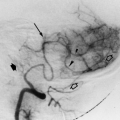
Angiographic Aspects
Long Axial View
In this view, the left border of the right atrium corresponds to the anterior portion of the interatrial septum, and the superior border is formed by the free superior wall of the right atrium. The lateral border is represented by a continuous line between the superior and inferior caval veins. The inferior border of the right atrium is the tricuspid valve and it overlaps the junction of the inferior vena cava. The anterior wall and the right auricle are not seen in this projection (Fig. 13.11).
Elongated Right Anterior Oblique View
The superior and inferior venae cavae are separated by a straight line that is the right posterior wall of the right atrium. The tricuspid valve seen in profile is in the inferior and left aspect of the atrial chamber. There is an inferior contour between the inferior vena cava and the tricuspid anulus, wherein is located the entrance of the coronary sinus and is formed partially by the atrioventricular septum. The left and superior aspect of the right atrium corresponds to the right atrial appendage or right auricle (Fig. 13.12).
Four-Chamber View
In this projection, the right atrium has a globular form, very similar to that seen in the long axial view. The left border corresponds to the more posterior portion of the atrial septum and the right border is related with the anterolateral atrial wall. The atrial appendage and the venae cavae are overlapped by the right atrium contour. The tricuspid valve is not well defined in this projection, although it forms the left inferior contour (Fig. 13.13).
Right Ventricle
Anatomic Aspects
The right ventricle is a triangular-shaped chamber and is located at the ventral portion of the heart. The base of the right ventricle is more cranial and to the right and the apex is caudal and projected toward the left. The base is formed at the right by the atrioventricular anulus and the leaflets of the tricuspid valve. At the left and more cranially is located the pulmonary valve. These two valves are separated by a smooth and prominent muscular invagination of the right ventricular wall called the ventriculoinfundibular fold. The rest of the right ventricle, including the apex, has a coarse trabeculation (Figs. 13.14, 13.15). The right ventricular chamber is divided into three portions: the inlet, the outlet, and the trabecular zones (Fig. 13.16). The inlet zone includes the tricuspid valve and extends until the implantation line of the papillary muscles. The outlet zone or infundibulum is a tubular muscular formation with the pulmonary valve on its top. The trabeculated zone extends from the papillary muscle’s insertion until the apex. The right ventricle is limited by three walls: the anterior or free wall, the inferior wall, and the septal wall, which corresponds
to the interventricular septum. The interventricular septum is formed by two components: the membranous septum and the muscular septum. The membranous septum is a small fibrous structure divided into two portions by the septal tricuspid leaflet attachment; the superior is the atrioventricular portion and the inferior is the interventricular portion. The atrioventricular portion is above the tricuspid anulus and separates the left ventricle from the right atrium. The interventricular portion is related to both ventricles. The muscular component, the largest part of the interventricular septum, is divided into three portions: the inlet portion, which divides the inlet of the ventricles; the infundibular portion, which separates the outlet of the ventricles; and the trabeculated portion, situated more apically. The outlet or infundibulum of the right ventricle is limited anteriorly by the free anterior ventricular wall. The posterior wall is the ventriculoinfundibular fold, the muscular formation that separates the tricuspid valve from the pulmonary valve. The third wall of the infundibulum is the infundibular or outlet portion of the interventricular septum. In normal hearts, the muscular structure, which separates the tricuspid from the pulmonary valve, is called supraventricular crest and is formed in its greater part by the ventriculoinfundibular fold and a small portion of the outlet septum. On the right side of the muscular interventricular septum, there is a well-marked muscular band called the septomarginal trabecula, which has two limbs embracing the body of the supraventricular crest. These three structures, the ventriculoinfundibular fold, the outlet septum, and the septomarginal trabecula, characterize the normal right ventricle (Fig. 13.17).
to the interventricular septum. The interventricular septum is formed by two components: the membranous septum and the muscular septum. The membranous septum is a small fibrous structure divided into two portions by the septal tricuspid leaflet attachment; the superior is the atrioventricular portion and the inferior is the interventricular portion. The atrioventricular portion is above the tricuspid anulus and separates the left ventricle from the right atrium. The interventricular portion is related to both ventricles. The muscular component, the largest part of the interventricular septum, is divided into three portions: the inlet portion, which divides the inlet of the ventricles; the infundibular portion, which separates the outlet of the ventricles; and the trabeculated portion, situated more apically. The outlet or infundibulum of the right ventricle is limited anteriorly by the free anterior ventricular wall. The posterior wall is the ventriculoinfundibular fold, the muscular formation that separates the tricuspid valve from the pulmonary valve. The third wall of the infundibulum is the infundibular or outlet portion of the interventricular septum. In normal hearts, the muscular structure, which separates the tricuspid from the pulmonary valve, is called supraventricular crest and is formed in its greater part by the ventriculoinfundibular fold and a small portion of the outlet septum. On the right side of the muscular interventricular septum, there is a well-marked muscular band called the septomarginal trabecula, which has two limbs embracing the body of the supraventricular crest. These three structures, the ventriculoinfundibular fold, the outlet septum, and the septomarginal trabecula, characterize the normal right ventricle (Fig. 13.17).
The tricuspid valve consists of an atrioventricular orifice surrounded by a fibrous ring, three somewhat triangular cusps or leaflets, various types of chordae tendineae, and papillary muscles. The cusps are named anterior, septal, and posterior. The anterior cusp is the largest and is interposed between the atrioventricular ring and the infundibulum. The septal cusp is attached to the membranous portion of the interventricular septum. The posterior cusp is attached in the inferior portion of the tricuspid annulus. The papillary muscles in the right ventricle are the anterior with the base arising from the anterolateral ventricular wall and related to the septomarginal trabecula, and the posterior, which is smaller than the anterior, arising from the inferior portion of the septum. There are small, papillary muscles arising from the infundibular septal wall.
The pulmonary valve is situated at the summit of the infundibulum. It consists of three semilunar segments or cusps attached to a fibrous annulus. Two of the cusps are anterior (right and left) and the third is posterior.
Angiographic Aspects
Long Axial View
In this view, the right ventricle has a triangular shape with the base at the top. The tricuspid valve is at the right and the pulmonary valve is at the left and in an upper level. The right contour corresponds to the free anterior wall. The upper left border is formed by the anterior portion of the interventricular septum and the posterior portion is a straight line toward the apex. The right ventricular outflow tract is limited by the supraventricular crest on the right side and by part of the septomarginal trabecula on the left. It looks like as a wide channel with the pulmonary valve on the top. The negative shadow of the tricuspid valve lies in the right and upper contour of the right ventricle. The anterior leaflet can be visualized superiorly and to the right on the tricuspid anulus. The septal leaflet is seen near and parallel to the interventricular septum. The posterior or mural leaflet is not visualized (Fig. 13.18).
Elongated Right Anterior Oblique View
The tricuspid valve seen in the lateral view is in the posterior border and to the right. The outflow tract is superior and to the left and is limited posteriorly by the supraventricular crest and anteriorly by the free wall of the right ventricle (Fig. 13.19).
Four-Chamber View
The morphologic aspect of the right ventricle in this projection is similar to that of the long axial view but the outflow tract is not visualized, and the tricuspid valve is localized more medially.
Left Atrium
Anatomic Aspects
The arterial blood returns from the lungs to the left heart through two pulmonary veins in each side of the left atrium. This is the most dorsal chamber and is localized in front of the lumbar spine and esophagus. It communicates with the left ventricle through the mitral valve. The left atrium has a quadrangular shape and a smooth posterior wall to which the four pulmonary veins converge. To the right, there is the interatrial septum and to the left there is an elongated pouch with a trabeculated wall that encircles the left aspect of the pulmonary artery; that is the left auricle or left atrial appendage. The left appendage is a finger-like formation that communicates with the left atrium through a narrow orifice. It is different from the right appendage, where the communication with the right atrium is wide and has a triangular form. The inferior wall of the left atrium corresponds to the mitral valve (Fig. 13.20).
Angiographic Aspects
Long Axial View
The right contour of the left atrium is formed by the anterior portion of the atrial septum. In the right upper corner, there is the entrance of the superior right pulmonary vein. The left pulmonary veins and the left atrial appendage are not seen. At the floor of the left atrium there is the mitral valve (Fig. 13.21).
Elongated Right Anterior Oblique View
In this view, the most prominent structure is the left atrial appendage, which forms the anterior and lateral borders of the left atrium. This is an irregular and elongated finger-shaped structure that protrudes toward the left between the superior wall and the mitral valve. The entrance of the right superior pulmonary
vein is localized on the right in continuation with the roof of the left atrium (Figs. 13.22, 13.23).
vein is localized on the right in continuation with the roof of the left atrium (Figs. 13.22, 13.23).
Four-Chamber View
This view shows an appearance similar to that described in the long axial view, but the atrial septum is visualized in its posterior portion (Fig. 13.24).
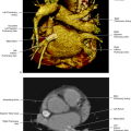
Computerized Tomographic (CT) Imaging
Computerized tomography is particularly useful for visualizing the left atrium and pulmonary veins because of the 3D volume images that can be generated. This 360° visualization of the pulmonary arteries can be used to rotate the image to quickly determine the number and size of vein, for instance (Fig. 13.25). The large size of the veins makes them easily identifiable.
Left Ventricle
Anatomic Aspects
The left ventricle has the thickest chamber wall of the heart and is located at the left and posterior to the right ventricle (Fig. 13.26). It has an elongated and triangular shape with the base upward where the mitral and aortic valves are located. The apex is oriented inferiorly toward the left. The left ventricle is divided into three portions: the inlet tract with the mitral valve complex, the trabeculated zone with less coarse trabeculation than in the right ventricle, and the outlet tract that supports the aortic valve. Unlike the right ventricle, in the left ventricle the inlet and the outlet tracts are not separated by a well-developed infundibulum and the aortic and mitral annuli are in continuity. The left ventricle has a lateral free wall, an inferior or diaphragmatic wall, and a septal wall. This one is a nontrabeculated wall that extends from the aortic annulus to the apex (Figs. 13.21, 13.27, 13.28, 13.29).
The mitral valve is formed by two leaflets or cusps attached to a fibrous anulus, a number of chordae tendineae, and two papillary muscles: the anterolateral and the posterior. The cusps or leaflets are the anterior or septal and the posterior or parietal, separated, one from the other, by two indentations: the anterolateral and the posteromedial commissures. The anterior cusp is longer and narrower than the posterior, but both have about the same area.
At the top of the left ventricular outlet tract is located the aortic valve, which consists of three cusps attached to the aortic anulus. Two cusps are posterior, right and left, and one is anterior. The anterior cusp is related to the right coronary artery above and to the membranous portion of the ventricular septum below, and is called the right coronary or septal cusp. The left posterior cusp is related to the left main coronary artery and is called left coronary cusp. The right posterior cusp is the noncoronary cusp.
Angiographic Aspects
Long Axial View
In this projection, the right contour of the left ventricle corresponds to the trabecular portion of the interventricular septum. A small cranial portion of the septum is formed by the outlet septum located just below the aortic valve. The outlet of the left ventricle is limited anteriorly and at the right by the outlet portion of the ventricular septum and posteriorly by the anterior leaflet of the mitral valve. The free wall of the left ventricle corresponds to the posterolateral contour and extends from the mitral valve to the apex. The mitral valve is seen as a negative shadow in the superior and lateral contour of the left ventricle. The papillary muscles are seen as a negative shadow in the middle portion of the left ventricle (Fig. 13.30).
Elongated Right Anterior View
In this view the outflow tract is limited anteriorly, and, to the left, by the infundibular septum as a straight vertical line below the right coronary cusp, and posteriorly, to the right, by a smooth contour extending from the noncoronary cusp to the crux cordis. It represents the atrioventricular portion of the interventricular septum. The anterior free wall of the left ventricle extends from the infundibular septum toward the apex, and the inferior wall corresponds to the contour from the crux cordis to the apex. The mitral valve is not well visualized in this view. The aortic valve is localized in the uppermost aspect of the outlet tract, and the coronary cusps cover each other at the left. The noncoronary cusp is at the right (Fig. 13.31).
Related posts:
Stay updated, free articles. Join our Telegram channel

Full access? Get Clinical Tree


