Arteries of the Spinal Cord and Spine
The arterial supply of the cervical spinal cord arises from several branches of the subclavian artery (Figs. 5.1, 5.2), vertebral artery (Fig. 5.3), deep cervical artery (Fig. 5.4), and ascending cervical artery, whereas the thoracolumbar segment is vascularized by branches of the thoracoabdominal aorta through the intercostal and lumbar arteries, and, occasionally, from the iliac and sacral arteries as well. The spinal cord has two almost independent arterial systems—or longitudinal anastomotic chains—one anterior and two posterior.
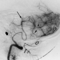
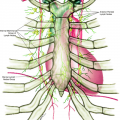
In the upper cervical region, the anterior spinal artery originates from the junction of the intradural segment of the vertebral arteries, just below the basilar artery (Fig. 5.5). In all other segments, the arteries cross through the intervertebral foramina to reach the intrathecal level. These are called segmental arteries (intercostal and lumbar arteries). The segmental artery divides into an anterior branch (along the costal groove) and a posterior branch to the spine. The posterior branch originates the muscular branches and the medial radiculomedullary artery. The radicular artery further bifurcates into two branches (the dorsal and ventral vertebral branches) and continues as the radicular artery and gives off a ganglionic branch and divides into an anterior spinal radicular artery and a posterior spinal radicular artery, following each anterior and posterior nerve root. In some preferential levels, these arteries are larger and constitute the anterior and posterior radiculomedullary arteries, proceeding as direct connections within the radicular artery and the longitudinal anterior and posterior anastomotic chains on the spinal cord surface (Fig. 5.6).
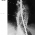
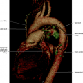
The anterior spinal artery is located in the midline on the ventral aspect of the cord, lying in the groove of the anterior median fissure of the spinal cord. It is formed by the union of two branches from the terminal portion of the vertebral artery at the level of the foramen magnum. The anterior spinal artery descends as a single trunk (Fig. 5.7) in the entire length of the ventral aspect of the medulla spinalis to the conus medullaris. It is reinforced by a succession of small spinal rami at the cervical level from the vertebral arteries, and by larger branches from the ascending cervical artery at the level C4–C6. Most of the tributaries in the lower two thirds of the cervical spinal cord are derived from the deep cervical artery at the level C6–T1. The superior intercostal artery may also send a branch to the spinal arteries. The anterior spinal artery continues downward, being reinforced by branches from the thoracic and abdominal aorta down to the conus medullaris, continuing along the cauda equina, and ending as a fine artery at the filum terminale. At that level, there are anastomoses with branches from the iliolumbar artery. The anterior radicular arteries, joining the anterior spinal artery vary considerably in size, number, and location. The total varies from 3 to 15, with an average of 7. The cervical region of the spinal cord receives an average of three arteries, the thoracic region has an average of three to four, and the lumbar region has an average of one. The position of the great anterior radicular artery, also called artery of Adamkiewicz, varies from T8 to L3 (Figs. 5.8, 5.9, 5.10), being often the only ventral feeder to the lower thoracic and lumbosacral cord (Fig. 5.11). The size of the anterior spinal artery usually tapers gradually from the lower part of the cervical region down to the middle or lower thoracic region, also narrowing in the upper thoracic region in some cases. At the point of anastomoses with the artery of Adamkiewicz, an enlargement of the anterior spinal artery usually occurs, remaining constant in size down to the lower end of sacral region, being reduced at that point to a tiny vessel, after giving off communicating branches, called rami cruciantes, to the posterior spinal arteries. This tiny artery may have anastomoses with branches of the iliolumbar artery.
Related posts:
Stay updated, free articles. Join our Telegram channel

Full access? Get Clinical Tree


