Cardiac Veins
The human heart has three systems of veins: the left ventricular, the right ventricular, and the thebesian veins. These are separate but intercommunicating systems.
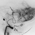
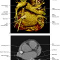
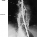
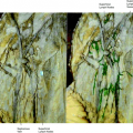
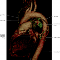
The left ventricular system drains most of the left ventricular venous blood and is formed by the anterior interventricular vein, the left marginal vein, the middle cardiac vein, and the right marginal vein (Fig. 14.1). The anterior interventricular vein ascends parallel to the left anterior descending (LAD) artery (Fig. 14.2) and enters into the left atrioventricular sulcus, where it becomes the great cardiac vein (Fig. 14.3). The great cardiac vein follows in continuity with the coronary sinus that ends in the right atrium (Fig. 14.4). The posterior ventricular vein, or middle cardiac vein, that runs in the posterior interventricular sulcus may drain into the right atrium or into the coronary sinus. The left marginal vein drains into the great cardiac vein (Fig. 14.5). The right marginal vein ends in the coronary sinus or in the right atrium (Fig. 14.6).
Related posts:
Stay updated, free articles. Join our Telegram channel

Full access? Get Clinical Tree


