Veins of the Thorax
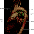
Brachiocephalic veins, also called innominate veins, are large veins at the upper thorax and represent united trunks of the internal jugular veins and subclavian veins. There are no valves in these veins (Figs. 8.1, 8.2, 8.3, 8.4, 8.5, 8.6).
The right brachiocephalic vein is approximately 2.5 cm long, following an almost vertical direction in front of the right brachiocephalic trunk, and joining the left brachiocephalic vein, thereby forming the superior vena cava. The tributaries are the right vertebral, internal thoracic, inferior thyroid, and occasionally the first right intercostal veins (Fig. 8.7).
The left brachiocephalic vein is 6 cm long, following an oblique path to the right direction to join the right brachiocephalic vein to form the superior vena cava. It is positioned anteriorly to the left subclavian and common carotid arteries. Tributaries are the left vertebral, internal thoracic, inferior thyroid, superior intercostal, thymic vein, and pericardiophrenic veins. Variations of the brachiocephalic veins include entering the right atrium separately and the configuration of a left vena cava.
Brachiocephalic Veins Internal Thoracic Veins (Mammary) (Fig. 8.8)
The internal thoracic veins are venae comitans to the internal thoracic arteries, ending in the corresponding brachiocephalic veins. Tributaries are the intercostal veins and pericardiophrenic veins.
Inferior Thyroid Veins (Fig. 8.9)
The inferior thyroid veins originate in the glandular venous plexus, having connections with the middle and superior thyroid veins. There is a pretracheal venous plexus, from which the left inferior vein descends to enter the left brachiocephalic vein, and the right inferior thyroid vein crosses the neck to open in the right brachiocephalic vein.
Left Superior Intercostal Vein (Fig. 8.1)
The left superior intercostal vein drains the second and third left posterior intercostal veins, directly to the left brachiocephalic vein.
Superior Vena Cava (Fig. 8.10)
The superior vena cava is the main vein for venous drainage for the superior aspect of the body. It is around 7 cm in length and is formed by the confluence of the brachiocephalic veins. It has no valves and ends in the right atrium. The superior vena cava is in contact with the right lung, pleura, trachea, right pulmonary hilum, and aorta. Tributaries are the azygos vein and small veins from the mediastinum.
Pericardiophrenic Vein (Fig. 8.11)
The pericardiophrenic veins are the main venous drainage channel for the diaphragm and the pericardium. These veins are in contact with the pericardium and pleura.
The thymic veins are small in adults, unless there is some enlargement of the thymus gland.
Azygos Vein
The azygos vein is formed by the confluence of the ascending lumbar veins, subcostal veins, and lumbar azygos. It ascends in the posterior mediastinum up to the level of the fourth vertebra, where it arches anteriorly above the right pulmonary hilum, ending in the superior vena cava. Tributaries are the posterior intercostal veins, the hemiazygos, the accessory hemiazygos veins, and the esophageal, mediastinal, and pericardial veins. The right bronchial veins also drain to the azygos vein, near the hilum. When present, the trunk formed by the subcostal and ascending lumbar veins is a major tributary of the azygos vein. The azygos vein starts laterally to the vertebral bodies but turns anterior to the thoracic spine as it approaches the vena cava (Figs. 8.15, 8.16, 8.17, 8.18, 8.19, 8.20).
Related posts:
Stay updated, free articles. Join our Telegram channel

Full access? Get Clinical Tree


