Pulmonary Venous Circulation
The pulmonary veins arise from the capillaries of the alveolar meshwork and from the capillary network of the pleura. The larger branches run within the interlobular septa and are, therefore, separate from the pulmonary bronchoarterial pathways. Histologically, the smaller pulmonary veins are identical to the arterioles. As the diameter of the veins increases, smooth muscle cells and elastic laminae are identifiable, and at a diameter of 60 and 100 μm, they become evident as veins and enter the interlobular septa (see Chapter 10, Fig. 10.18).
There is a dense vascular network around the bronchi, which is an arteriolar network terminating in bronchial capillaries and numerous bronchial venous plexuses with a characteristic irregular shape and course. There are also connections between the bronchial capillaries with the bronchial venous plexuses. The bronchial venous plexus communicates with the smaller branches of the pulmonary vein.
Pulmonary Veins
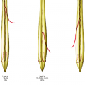
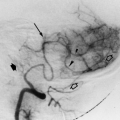
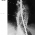
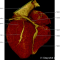
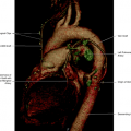
There are usually two superior and two inferior main pulmonary veins, the former draining the middle and upper lobes on the right side and the upper lobe on the left with the inferior veins draining the lower lobes (Fig. 11.1). The pulmonary veins drain the oxygenated pulmonary blood to the left atrium. Computerized tomography is now used as an excellent way to image these structures (Fig. 11.2). As with many venous structures, there are occasional variations in this pattern. On the right side the most common variation is to have three separate pulmonary veins with the third vein usually draining the right middle lobe. On the left side the most common variation is a single common trunk that all the veins empty into before reaching the left atrium (Fig. 11.3). Less commonly more complex arrangements may be seen (Fig. 11.4). These patterns are usually referred to as normal variants, since the blood still follows the same pathway.
In anomalous pulmonary venous drainage, the veins drain into a different structure, usually directly to the superior vena cava or to the right atrium. This will be discussed in more detail in subsequent text.
The right veins reach the hilum beneath the main pulmonary artery and posteriorly to the superior vena cava. The veins usually reach the left atrium independently as two separate veins. The left veins cross in front of the descending aorta and may enter the left atrium separately as on the right side or may join each other already within the pericardial cavity to enter the atrium as a common vein (Figs. 11.5, 11.6, 11.7, 11.8).
Related posts:
Stay updated, free articles. Join our Telegram channel

Full access? Get Clinical Tree


