Lymphatic System of the Thorax
Peripheral lymph drainage to the venous circulation is accomplished via the right and left lymphovenous portals, which are located close to the junction of the large internal jugular and subclavian veins. On the right, there are three main lymphatic trunks; on the left, four main lymphatic trunks are identified: the three corresponding to the right and in addition the thoracic duct, which is the largest lymphatic trunk in the body (Figs. 9.1, 9.2, 9.3, 9.4, 9.5). The anatomy of the lymphovenous portals varies. In 80% of subjects, the right trunks open independently at the jugular–subclavian junction. In one fifth of cases, a short right lymphatic trunk is formed.
Thoracic Lymphatic Drainage
Lymphovenous Portals
On the Right
There are three main lymphatic trunks converging to the junction of the right internal jugular vein with the right subclavian vein: (1) the right jugular trunk, which runs along the ventrolateral aspect of the internal jugular vein, carrying all the lymph from the right half of the head and neck; (2) the right subclavian trunk from the right apical axillary lymph nodes, running along the axillary and subclavian vein, and carrying lymph from the upper right limb and the superficial tissues of the right chest and abdominal wall; and (3) the right bronchomediastinal trunk, which runs along the trachea, carrying lymph from the right lung, diaphragm, bronchi, and trachea.
On the Left
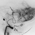
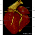
The left portal receives the volume of lymph from all over the body, except the territories mentioned for the right lymphatic trunks (Figs. 9.6, 9.7, 9.8). There are four main lymphatic trunks converging to the left lymphovenous portal: (1) the left jugular trunk, which runs along the ventrolateral aspect of the internal jugular vein, carrying lymph from the left half of the head and neck; (2) the left subclavian trunk draining lymph from the upper left limb and superficial tissues of the left chest and abdominal wall; (3) the left bronchomediastinal trunk, which drains more of the heart and esophagus, in addition to the lung, bronchi, and trachea; and (4) the thoracic duct, which drains the remaining territories of the body. It is formed by the abdominal confluence of the lymphatic trunks to the cisterna chyli.
Thoracic Duct
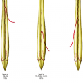
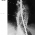
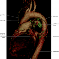
The thoracic duct measures about 38 to 45 cm in length, in adults, extending from the abdomen to the neck, transgressing the diaphragm, ascending at the posterior mediastinum right of the midline, between the descending thoracic aorta and the azygos vein and anterior of the vertebral column. The thoracic duct commences wide, forming in some cases a pouch called cisterna chyli, and diminishes in caliber up to the neck, where it arches laterally to the left and anteriorly, and eventually descends in a direction to the junction of the left internal jugular and left subclavian veins, opening into the venous system through a bicuspid valve. At the termination, the thoracic duct may be multiple, and the site or opening may be in any of the left great veins. The origin of the thoracic duct is the confluence of the four main abdominal lymph trunks (Figs. 9.1, 9.2, 9.3, 9.5).
Related posts:
Stay updated, free articles. Join our Telegram channel

Full access? Get Clinical Tree


