Introduction
Although a comprehensive hip examination will be described, in the majority of cases the examination is focused on the particular presenting symptom. Potential causes of anterior hip pain extend from the pubic symphysis to the hip joint itself. Patients with lateral hip pain most frequently require an examination of the gluteal insertion and associated bursae. Posterior pain could be due to abnormalities extending from the hamstring attachment to the ischium to the posterior aspect of the hip joint.
Position 1: Adductor Insertion
Imaging Goals
- 1.
Identify the common adductor tendon and its relationship to the pubic symphysis.
- 2.
Locate the muscle bellies of the adductor compartment.
- 3.
Locate gracilis.
Technique
The patient lies supine with the hip and knee flexed and the hip externally rotated into the frog leg position.
| The superficial and most palpable muscle on the medial thigh is the adductor longus. |


The probe is then turned into the axial plane and moved slightly towards the opposite side to reveal the anterior margin of pubic symphysis. The integrity of the anterior annulus and the bony contour of the adjacent pubic bones can be assessed.
A prominent pubis or irregular bony surface may suggest symphysitis, though similar changes are found in asymptomatic individuals.
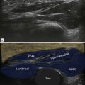
From the common tendon insertion, the probe is moved distally until the three adductor muscles are found. From anterior to posterior they are: adductor longus, brevis and magnus. The common adductor tendon has usually split by this point, the larger anterior component being the tendon of adductor longus. Just behind this, the smaller component opens into a fourth muscle that overlies the adductors, the gracilis muscle ( Fig. 16.3 ).

Position 2: Anterior Hip Joint
Imaging Goals
- 1.
Locate hip joint.
- 2.
Locate the iliopsoas tendon.
- 3.
Locate the obturator nerve.
Technique
Returning to the level of the adductor tendon, the axially positioned probe is moved laterally along the superior pubic ramus. Pectineus is the muscle that separates the probe from the underlying pubic bone. Further lateral movement reveals the anterior acetabular wall, anterior labrum and rounded contour of the femoral head ( Fig. 16.4 ). A small quantity of fluid may be detected in the hip joint. The psoas muscle/tendon lies lateral to pectineus, with the fleshy iliacus component distinguishable from the tendinous psoas portion. A little more distally the vastus intermedius appears in this position.
Abnormal iliopsoas tendon movement may underlie the clicking sensation felt by some patients with snapping hips.
| Iliopsoas snap can be elicited by abducting, flexing and externally rotating the hip and then bringing it back to the normal neutral position. |

Returning to pectineus, the probe can be moved a little distally to just below superior pubic ramus. Once the bone falls away from view, a neurovascular bundle is identified just beneath pectineus, surrounded by a small cuff fat ( Fig. 16.5 ). This is the obturator nerve and artery. It is easy to find in thin people, but becomes rather deep and difficult to locate with enlarging body habitus. Moving the probe further distally still, the upper part of the adductor longus is seen with the other adductor muscles and the obturator externus below. If the probe is moved a little laterally, the bony contour of the femur is found and can be tracked distally until the lesser trochanter is visualized. This is a good position to identify the insertion of the iliopsoas tendon; some rotation of the probe is required to orientate the tendon in long axis.

On the anterior aspect of the hip, the anterosuperior labrum, anterior capsule and femoral head articular cartilage are noted.
| The degree of joint effusion is best assessed at the head–neck junction. |
Position 3: Sartorius, Abdominal Wall and Nerves
Imaging Goals
- 1.
Identify the sartorius origin.
- 2.
Locate the abdominal wall muscles.
- 3.
Locate the ilioinguinal and lateral cutaneous nerves.
Technique
The area above the hip joint is now examined. The anterior margin of the iliac bone is followed upwards to the anterior superior iliac spine (ASIS). The oval-shaped tendon of sartorius can be seen to arise from it. Medial to this are the muscles of the abdominal wall. These form three layers; from superficial they are: obturator externus, obturator internus and transversus abdominis ( Fig. 16.6 ). The ilioinguinal nerve can be located between the second and third muscle layers. At the lower margin of the abdominal wall, a ligamentous structure passes from the ASIS to a small tubercle on the pubic bone. This is the inguinal ligament. The lateral cutaneous nerve of the thigh lies beneath it, close to its lateral attachment, surrounded by a small cuff of fat. The nerve passes distally on the superior surface of sartorius. The attachment of sartorius itself is better assessed by rotating the probe 90° to view it in long axis. Lateral to the ASIS is the origin of the iliotibial band.


