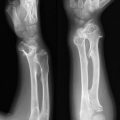
Localization: It is generally localized in the metaphysis at first and then displaced toward the diaphysis. It starts intracortically/subperiosteally. Most of the lesions are located around the knee and ankle. It is rare in the proximal femur and upper limb, exceptional in the trunk, hand, and foot. Two or three lesions may appear in the same or both lower limbs (e.g., distal femur and proximal tibia).
Clinical Aspects: Diagnosis is usually based on a radiogram obtained for unrelated reasons (usually trauma), histiocytic fibroma being the most classic and common lesion diagnosed as incidental finding. Rarely, pathologic fractures may occur in larger lesions (more than 1/2–2/3 of the bone cross section, particularly for the distal tibia and fibula).
Imaging: Standard X-rays are usually diagnostic. The defect is metaphyseal, intracortical, and/or subperiosteal. Generally lobulated, its inner boundaries are surrounded by a rim of bone sclerosis. The osteolytic image may appear multilocular due to corrugations of the bony wall. The cortex is sometimes attenuated, rarely slightly expanded due to chronic periosteal reaction.
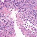
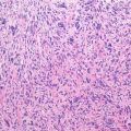
Pathology: Tissue is compact, rubbery soft, tan-brown in color, sometimes containing yellow (foam cells) or dark (hemosiderin) areas. Dense network of cellular typically plump spindle cells with a prominent storiform pattern and with scattered multinucleated giant cells are evident. Intra- and extra-cytoplasmatic hemosiderin and lipid-loaded foam cells are common findings.
Course and Staging: Tumor growth stops at skeletal maturity and very frequently even before. The lesion then tends to ossify slowly. Stage is initially 1 or 2 and then constantly becomes 1.
Treatment and Prognosis: The majority of histiocytic fibromas do not require treatment; diagnosis can usually be made on clinico-radiographic features avoiding biopsy. Occurrence of a pathologic fracture per se is not an indication for surgery, as fractures heal essentially in a normal fashion. Curettage and bone grafting are rarely needed for large lesions, possibly associated with internal fixation for displaced fractures.
Key Points
Clinical
Related posts:Stay updated, free articles. Join our Telegram channel
Full access? Get Clinical Tree
 Get Clinical Tree app for offline access

|