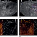
Rintaro Hashimoto and Kenneth J. Chang University of California, Irvine, Orange, CA, USA Portal hypertension (PH) is a serious adverse event of liver cirrhosis. Clinically significant PH (>10 mmHg) predicts the development of esophageal varices bleeding and hepatocellular carcinoma. Portal venous pressure (PVP) accurately reflects the degree of PH and is the single best prognostic indicator in liver disease. The evaluation of PVP had been performed by an indirect measurement of the hepatic venous pressure gradient (HVPG), in which a catheter is inserted into the hepatic vein percutaneously via either the jugular or femoral vein. This technique requires skill and is not commonly performed. HVPG can be affected by hepatic tissue compliance. In addition, HVPG has been found to differ from PVP even among patients with the same pathogenesis. Recently, a simple novel technique for EUS‐guided portal pressure gradient measurement (PPGM) using a compact manometer (Cook Medical, Bloomington, IN) (Figure 40.1) has been developed. This enables measurement of portal venous pressure with the manometer directly through the endoscopic ultrasound (EUS) needle. The time required for the procedure is short, under 30 minutes. EUS‐guided liver biopsies can be performed at the same procedure. The EUS manometry apparatus used in our human study is a simple set‐up that includes a 25‐gauge fine needle aspiration (FNA) needle, noncompressible tubing, a compact digital manometer, and heparinized saline. The tubing is connected by a Luer lock to the distal port of the manometer while the heparinized saline is connected the proximal port. The end of the tubing is connected via a Luer lock to the inlet of the 25G needle. The patient is positioned supine and during EUS‐guided pressure measurement reading, the manometer is placed at the patient’s mid‐axillary line (Figure 40.2). We prefer monitored anesthesia care or general anesthesia for this procedure. The hepatic venous pressure is measured first. Of the hepatic veins, the middle hepatic vein is targeted most commonly because of its larger caliber and better alignment with the needle trajectory on linear EUS (Figure 40.3). Doppler flow is used to confirm the typical multiphasic waveform of hepatic venous flow (Figure 40.4
40
How to do Endoscopic Ultrasound‐guided Portal Pressure Gradient Measurement
Introduction
Endoscopic ultrasound‐guided PPGM technique
![]()
Stay updated, free articles. Join our Telegram channel

Full access? Get Clinical Tree


