Fig. 26.1
Adult Echinococcus granulosus
26.1.1.2 Egg
The egg of E.g. resembles to the egg of Taenia saginata parasitic to cows and pigs. They are wide oval in a color of yellowish brown, with two layers of embryonic membrane and inner radiating ridge (Fig. 26.2). After maturation, just before or after discharge of the gravid proglottid via the intestinal canal of the host, its uterine ruptures to discharge eggs. The eggs have strong resistance to external environment, which can survive for above 2.5 years in water at 2 °C. However, the eggs are heat intolerant and can be killed at a temperature of 80 °C. The eggs can survive for 6–24 h in common disinfectants and chemical agents, such as 20 % Lysol, and can survive for 24 h in 20 % formaldehyde.
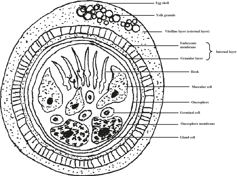

Fig. 26.2
An egg of Echinococcus granulosus
26.1.1.3 Larva
The larva of E.g. is echinococcus, which is a round or round-like cystid. The cystid wall is composed of an external transparent cuticle and an internal germinal layer, which is enveloped by a fibrous capsule formed by host tissue response (Fig. 26.3). The germinal layer is embryonic membrane tissue capable of reproduction, with its lining capable of budding many small projections. These projections gradually develop into germinal cysts and then daughter cysts after shedding. In the daughter cysts, several scolices can be produced, which are known as protoscolices. In the daughter cysts, the granddaughter cysts can be produced. The cyst is filled with hydatid fluid. The size of echinococcus is related to the parasitic time, parasitic position, and host, which ranges from less than 1 cm to more than 10 cm. The large echinococcus contains more than 1,000 ml hydatid fluid. And echinococcus can survive in the body of its host for several years or even 20 years.
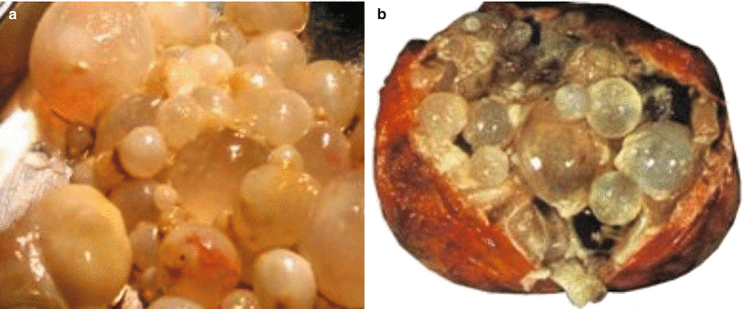

Fig. 26.3
Specimens of Echinococcus granulosus
26.1.2 Echinococcus multilocularis (E.m.)
26.1.2.1 Adult Worm
The adult worm resembles to that of E.g. but with an even smaller size (1.2–4.5 mm). The size of its egg is (30–38) μm × (29–34) μm.
26.1.2.2 Egg
The shape and size of E.m. eggs are similar to those of E.g., presenting difficulties for their differentiation.
26.1.2.3 Larva
E.m. is commonly parasitic in the liver and sometimes in the lungs and brain. Generally, it is a single giant vesicular mass like in the color of light yellowish or whitish. Sometimes, it is nodular. Otherwise, vesicular and nodular masses concur. E.m. reproduces in ways of budding or infiltration, with continuous production of new vesicles to grow into the tissues like neoplasms. It mostly grows outward, with rare occurrence of inward growth to form septum. Therefore, the cystid is separated into multiple small cysts. The new vesicles may invade the vascular or lymphatic vessels to metastasize into the lungs, brain, spleen, kidney, adrenal gland, heart, and even occasionally portal lymph nodes. Therefore, by naked eyes, the conditions may be misdiagnosed as hepatic carcinoma. Generally within 1–2 years, the parasitized organs are occupied by vesicles in different sizes. The grape cluster-like vesicles may spread from the organ surface into body cavities, which appear like carcinoma. Therefore, it is referred to as worm cancer.
26.2 Epidemiology
26.2.1 The Source of Infection
House dogs are the terminal host of E.g., which is also the most important source of infection. The adult worms parasitic in the intestines of dogs experience maturation of eggs and shedding of gravid proglottid in a cycle of 7–14 days. Continual egg excretion can be found in the feces of infected dogs.
26.2.2 Route of Transmission
Due to close contacts to dogs, the eggs of E.g. infect humans via contaminated hands and oral intake. The worm eggs in dog feces may contaminate vegetables and water sources, especially sharing water source in humans and animals, which also causes infection.
26.2.3 Susceptible Populations
The susceptible populations are mainly farmers, peasants, and the fur workers. However, because increasingly more people raise dogs in urban areas, the urban residents are also at high risk of infection. Most patients are infected during childhood and show the obvious symptoms after they develop into young or middle-aged adults.
26.2.4 Epidemiological Features
Cyst echinococcosis is distributed worldwide, but is more common in countries and regions with dominant industry of stockbreeding such as Australia, New Zealand, Argentina, Uruguay, South Africa, and Asia. In China, cyst echinococcosis mainly prevails in northwestern farming areas such as Xinjiang Wei Autonomous Region, Ningxia Hui Autonomous Region, Tibet, and Gansu, with sporadic cases in other regions.
Alveolar echinococcosis is sporadic, which is mainly distributed in Middle and South Europe, North America, and Britain. In China, the cases are reported in Qinghai, Ningxia Hui Autonomous Region, and Xinjiang Wei Autonomous Region.
26.3 Pathogenesis and Pathological Changes
26.3.1 Pathogenesis
After the eggs of E.g. enter the human body along with food intake, they are incubated into oncospheres under the effects of digestive juice. Some oncospheres invading into the tissues are surrounded by local cells and eliminated, with some other oncospheres surviving and developing. The oncospheres firstly invade the liver along with blood flow in the portal vein, with most forming hydatid cysts in the liver and rarely others invading the lungs via the hepatic sinusoid, hepatic vein, and right heart. The cases with following invasion of hydatid cysts into the systemic circulation via pulmonary microvascular vessels and the left heart show systemic involvements of organs. Therefore, it has been known that hydatid can be parasitic at any part of human body. The infective rate of each organ is directly related to the blood flow volume of each organ and the sequence of oncosphere invasion along with blood flow. Therefore, the organ with the highest to the lowest incidence rate is sequentially the liver, lungs, abdominal cavity, pelvic cavity, spleen, kidneys, and brain. The pathogenesis of CE is predominantly mechanic compression, and the other etiological factor is rupture of E.g. cyst-induced homologous protein allergy. E.g. grows very slowly and it usually lasts for about 10 years from infection to onset of symptoms.
After intake of food contaminated by worm eggs, oncospheres are inoculated in the intestines of humans, which then penetrate intestinal mucosa into the portal vein. After their arrival at the liver, they develop into hydatid.
26.3.2 Pathological Changes
The pathological changes of hydatidosis include space-occupying growth of the cysts and the consequent compression to the adjacent organs. E.g. gradually grows and matures in the liver, with intrahepatic cholangioles compressed and imbedded into the external cyst wall. Sometimes, the cholangioles penetrates into the cystic cavity due to compressive necrosis to stain the daughter cysts and the cyst fluid into yellow and to cause secondary bacterial infection. The hydatid cysts of lungs can penetrate into the bronchi. Occasionally, the germinal layer, scolex, and cyst fluid can be coughed up altogether, with complication of bacterial infection. If the hydatid cyst penetrates into the bronchioles, air fills in the space between internal and external cyst to show crescent air-containing area. If hydatid cysts of the liver rupture with the fluid flowing into the abdominal cavity, allergic shock may occur. The access of scolex into the abdominal cavity can cause multiple hydatidosis.
E.m. is in multilocular growth, with virtually all of its primary focus originating from the liver, and the foci in other organs are metastatic from the liver. The liver lesions are scattered light grayish-white nodules in different sizes on the liver surface, which are hardened with no envelope and poorly defined boundaries with surrounding liver tissues. On the cross sections of the lesions, there are necrotic tissues and cavities. Microscopy demonstrates vesicles in irregular shapes and different sizes, which are surrounded by infiltration of eosinophils granulocytes, lymphocytes, plasmocytes, and other inflammatory cells. E.m. surviving for several years is commonly surrounded by a thick layer of fibers, with no or small quantity of fluid in the cyst. At the cystic wall, there are calcium depositions, which are granular in amorphous calcification.
26.3.3 Types of Hydatidosis
26.3.3.1 Singular and Multiple Hydatidosis
Singular hydatidosis refers to infection of only one hydatid. Multiple hydatidosis refers to concurrent infection of at least two hydatids in one or more organs.
26.3.3.2 Primary and Secondary Hydatidosis
Primary hydatidosis is the infection of E.g. eggs, which grow and develop parasitically in host. Primary hydatidosis accounts for most of the hydatidosis cases. Secondary hydatidosis is infection of newly developed hydatid from transplanting germinal cyst, daughter cyst, or protoscolex as well as from asexual reproduction of primary hydatid cyst.
26.3.3.3 In Situ Relapse of Hydatidosis
After surgical removal of the hydatid, the residual daughter cyst or protoscolex in the external cystic cavity continues its growth into new hydatid that causes in situ relapse of hydatidosis.
26.3.3.4 Recurrent Hydatidosis
After hydatidosis is healed, a repeated infection of hydatid eggs causes hydatidosis again, which is known as recurrent hydatidosis.
26.4 Clinical Symptoms and Signs
In the early stage of the disease, the patients may have no subjective symptoms. The clinical manifestations are related to the parasitic site, size, and quantity of echinococcus as well as host responses and complications. Hydatidosis has a long incubation period, commonly 10–20 years or even longer from the infection to the onset of symptoms.
26.4.1 Hepatic Hydatidosis
26.4.1.1 Hepatic Cystic Hydatidosis
This type is the most common, with lesions commonly located in the right liver lobe. The patients may experience hepatic upset, dull pain or distended pain, enlarged liver, and protruding liver surface. Painless mass can be palpated that has smooth surface and is mobile along with breathing. By percussion, the mass feels trembling, which is known as hydatid thrill sign. The hydatid adjacent to hepatic hilum can compress the bile ducts to show jaundice. It can also compress the portal vein to cause portal hypertension. In the cases with complicated infections, lesions resembling to hepatic abscesses can be found. And its penetrating into abdominal cavity can cause diffuse peritonitis and allergic responses. In some serious cases, allergic shock occurs. Secondary abdominal or thoracic hydatidosis may occur due to dissemination of hydatid into abdominal or thoracic cavity along with cystic fluid.
26.4.1.2 Hepatic Alveolar Hydatidosis
In its early stage, the patients experience no upset. Along with progress of the conditions, the patients experience hepatic pain, poor appetite, abdominal distension, and other symptoms. In its late stage, the patients experience jaundice, cachexia, itchy skin, ascites, and other signs of portal hypertension. By physical examination, hardened painless mass can be palpated, which has smooth or nodular surface and tends to be misdiagnosed as hepatic carcinoma.
26.4.2 Pulmonary Hydatidosis
Pulmonary hydatidosis commonly occurs in the right lung, especially middle and lower lobes of the right lung. In the early stage, the patients commonly have no subjective symptoms and its early detection depends on physical examinations. The patients may complain of chest dull pain or stabbing pain, irritating cough, and hemoptysis. In the cases with hydatid cyst penetrating into the bronchus, the patients experience sudden dyspnea and paroxysmal cough with large quantity of watery cystic fluid along with sheet jellylike cystic wall and cystic sands. Occasionally, suffocation occurs due to overflow of cystic fluid and occlusion. In the cases with secondary infection, the patients experience high fever, chest pain, and cough with thick sputum.
26.4.3 Brain Hydatidosis
Its incidence rate is low, with more common occurrence in children. The lesion is commonly located at the parietal lobe, with accompanying hepatic and/or pulmonary hydatidosis. The clinical symptoms include headache, papilledema, and other symptoms of intracranial hypertension, with accompanying epilepsy attacks.
26.4.4 Spleen Hydatidosis
The patients experience falling pain, distension, and upset of the left epigastrium. The lesion commonly compresses the gastric fundus and the greater curvature of the stomach to cause abdominal distension, nausea, hiccups, poor appetite, and other digestive symptoms after food intake. However, complications rarely occur. The typical cases have palpable painless half sphere-shaped tough mass at the left epigastrium, which has smooth surface and feels elastic by pressure touch. By percussion, hydatid thrill sign can be found.
26.4.5 Kidney Hydatidosis
The patients experience dull pain of the lower back with falling pain, distension, and upset. The patients with long illness course commonly pay their clinic visit due to touchable painless mass at the epigastrium or lower back.
26.4.6 Abdominal and Pelvic Cavity Hydatidosis
Abdominal and pelvic hydatidosis can be divided into primary and secondary, with secondary cases being more common, which is secondary to rupture of hepatic hydatid or surgeries removing hepatic hydatid. Clinically, the common symptoms include abdominal distension, abdominal falling pain, poor appetite, and emaciation. The lesion compresses the intestinal canal to produce mechanical incomplete intestinal obstruction. The lesion compresses the diaphragm to elevate it and confine its movements, with consequent occurrence of shortness of breath and inability of supine position. The lesion may also compress the inferior vena cava to cause edema of lower limbs. Pelvic hydatid may compress the rectum and bladder to cause symptoms during defecation or urination. Hydatid in female reproductive organs may invade and compress the uterine and/or fallopian tubes to cause falling pain and distension of the lower abdomen as well as dysmenorrheal. After pregnancy, the hydatid can also be compressed with complicated hydatidosis by rupture of hydatid or infection.
26.4.7 Bone Hydatidosis
The patients may have painless mass or only local soreness and pain. The conditions tend to be misdiagnosed as chronic pain of soft tissues.
26.4.8 Cardiac Hydatidosis
Cardiac hydatidosis rarely occurs, with hydatid commonly parasitic in the left ventricle (accounting for about 50–60 % of cardiac hydatidosis cases). Other parasitic positions include the interventricular septum, right ventricle, pericardium, and left and right atrium according to the respective incidence rate. The parasitic position of cardiac hydatid is related to the route of infection and the blood supply of each part. In the early stage, X-ray commonly detects confined protrusion at the cardiac margin. With progress of the conditions, the patients experience symptoms of chest distress, palpitation, and arrhythmia.
26.4.9 Hydatidosis of Other Body Parts
Hydatidosis can also be found at the eye socket, mammary gland, subcutaneous tissues, muscles, pancreas, and thyroid gland.
26.5 Hydatidosis-Related Complications
26.5.1 Complications of Hepatic Hydatidosis
26.5.1.1 Hepatic Cyst Rupture
Hepatic hydatid can penetrate into the abdominal cavity to cause sudden severe abdominal pain and sudden shrinkage or disappearance of abdominal mass, with accompanying itchy skin, urticaria, dyspnea, cyanosis, vomiting, diarrhea, and other allergic responses. In some serious cases, shock and even death occur.
26.5.1.2 Secondary Infection
Secondary infection is commonly caused by penetration of the cyst into the bile duct. Clinically, its manifestations resemble to bacterial hepatic cyst and cholelithiasis. Due to ruptured cystic membrane or occlusion of daughter cyst, secondary infection occurs to cause purulent angiocholitis.
26.5.2 Complications of Pulmonary Hydatidosis
The incidence rate of secondary infection is 10–15 %, with concurrent infection and rupture, which are mutually cause and effect. Secondary infection occurs due to rupture. Occasionally, patients have no case history of rupture symptoms before the infection. Therefore, the infection is primary. After infection, hydatid cyst-bronchus fistula forms. Regardless of the formation of bronchial fistula, symptoms of pulmonary abscess occur, including chest pain, fever, emaciation, cough, and purulent sputum, which persist for a long period of time. In the cases with accompanying bronchial fistula, the patients experience coughing up purulent sputum with daughter cyst or internal cyst fragments and no hemoptysis when infection is not serious. When the bronchial fistula is smoothly drained, more purulent sputum is coughed up, followed by a decrease of body temperature and alleviated symptoms.
26.5.3 Complications of Abdominal Hydatidosis
26.5.3.1 Intestinal Obstruction
During the development of parasitic hydatid in the host abdominal cavity, the parasitized organ show fibrous tissue hyperplasia to surround the hydatid. Therefore, the external cyst is formed and its surrounding serous tissues response to exudate fibrin to form membranous or cord-like adhesion. Mechanical incomplete intestinal obstruction is resulted in, which may repeatedly occur with aggravated conditions.
26.5.3.2 Abdominal Infection
Its incidence rate is lower than that of hepatic hydatidosis, accounting for about 10 % of the cases with abdominal hydatidosis. It mainly occurs in the cases with multiple hydatidosis and a long history of illness. The external cyst of abdominal hydatid is thick and plays a role of barrier to confine the inflammation within the cyst. Therefore, the systemic inflammation is mild and the patients experience only dull pain, long-term low-grade fever, poor appetite, anemia, and emaciation.
26.6 Diagnostic Examinations
26.6.1 Laboratory Tests
26.6.1.1 Casoni Test
The operational procedures are simple and the test results can be obtained within 15 min. Casoni test has a positive rate of 68–100 %, which can be applied for primary screening.
26.6.1.2 Indirect Hemagglutination Test (IHA)
The operational procedures are quite simple and the test has a high specificity. It has a positive rate of about 82 % for hepatic hydatidosis.
26.6.1.3 Enzyme-Linked Immunosorbent Assay (ELISA)
Its sensitivity and specificity exceed IHA, with rare false-positive results.
26.6.1.4 Dot-ELISA
Its operational procedures are simple and the test results are directly observable. And the test is applicable in community-based hospitals.
26.6.1.5 ABC-ELISA
The sensitivity is the highest in these laboratory tests, being about four to six times as high as routine ELISA.
26.6.1.6 Detection of Specific Antibody of Hydatidosis
In April 2011, the specific antibody test kit for the diagnosis of hydatidosis was successfully developed in Xinjiang Medical University, China. The kit is for the diagnosis of hydatidosis with advantages of being fast, simple, specific, and accurate. It can be applied both in clinical diagnostic trials and field epidemiological investigations, functioning in both diagnosis and differential diagnosis (between CE and AE). The kit is the first serologic test for the diagnosis of hydatidosis, which standardizes the diagnostic criteria of hydatidosis. It is a favorable diagnostic tool for early diagnosis, differential diagnosis, and epidemiological studies of hydatidosis.
26.6.2 Radiological Examinations
26.6.2.1 Ultrasound
Ultrasound examination is a noninvasive and safe radiological technology, which renders direct observation for diagnosis and assessment of the diseases. It can clearly demonstrate the space-occupying lesion of hydatidosis, which provides repeated and dynamic observation of the lesions and can be operated conveniently.
26.6.2.2 X-Ray Radiology
X-ray radiology is the examination of choice for the diagnosis of pulmonary hydatidosis and bone hydatidosis. Due to the distinct contrast of density between lungs and bone tissues to hydatid cysts, their respective locations, quantity, size, properties, and complications can be well defined by X-ray.
26.6.2.3 CT Scanning
CT is an important examination for the diagnosis of hydatidosis, which is applicable to all organs of human body.
26.6.2.4 MR Imaging
For the diagnosis of complex hydatidosis, MR imaging is superior, especially magnetic resonance hydrography and magnetic resonance angiography which play important roles in the diagnosis of hydatidosis.
26.7 Imaging Demonstrations
26.7.1 Hepatic Hydatidosis
26.7.1.1 Hepatic Cystic Hydatidosis
Ultrasound
Based on the natural development of hepatic hydatid, different stages of pathological changes, and pathological changes of various complications as well as their sonogram, hepatic cystic hydatidosis can be divided into seven types.
Singular Type
Because the hydatid cyst is filled with watery cystic fluid, both real-time linear array and fan-shaped scanning demonstrate round or oval singular liquid dark area with no echo. However, small hydatid cysts have less tension and some of them may fail to show a round lesion area (Fig. 26.4). In the cases with the hydatid cyst located inferior to the hepatic capsule to form hump-like change, the cyst may compress the intrahepatic duct system and the adjacent right kidney and gallbladder. The smooth and thick hydatid cystic wall has obvious acoustic impedance different from hepatic parenchyma to form bright boundary. The large hydatid cysts has double-layered cystic wall (internal cystic wall and external cystic wall) that produces strong echo, which shows well-defined bright boundary from the organ tissues. The hydatid cystic wall is far thicker than other cysts. The ultrasound is reflected by the lateral margin of thick cystic wall to produce acoustic shadow at the lateral wall. Ultrasound demonstrates no echo area or no reflection area for liquid-contained hydatid cyst. Its demonstration depends on the potential space between the internal cyst wall and the external cyst wall as well as small quantity of liquid. Therefore, a fine dark interface is commonly demonstrated to show double-wall sign, which is characteristic at sonogram of hydatidosis. The ultrasound bundle produces acoustic energy via hydatid cystic fluid to increase the adduction, which shows the enhancement effect of the posterior wall of hydatid cyst. When the probe pressed, the hydatid cyst may be deformed. It should be cautious to avoid rupture of the hydatid cyst. When the hydatid cyst is vibrated with the ultrasound probe, there are floating fine light spots posterior to the cyst, which are stirred protoscolex to show reflected light spots that deposited at inferior to the cyst, which are known as cystic sands.
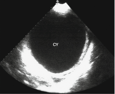

Fig. 26.4
Singular type of hepatic cystic hydatidosis. Ultrasound demonstrates liquid dark area in the left liver lobe (CY), with space between lateral wall of the internal cyst and external cyst and posterior enhancement effect
Multiple Type
In the liver, at least two round or oval cysts are probed, which are in different sizes but with characteristic sonogram of hydatid and septum of liver tissues or cystic wall of the hydatid.
Daughter Cyst Type
In the dark liquid area of the mother cyst, there are sphere-shaped dark light rings in different sizes and different quantities, namely, characteristic cyst-in-cyst sign of hydatidosis. In the cases with rare daughter cysts, the daughter cysts are arranged close to the mother cyst wall. In the cases with more daughter cysts, the mother cyst can be filled up, with the daughter cysts clinging and overlapping to form large quantity of small round light rings or dense short light strips, namely, honeycomb sign or grape cluster sign. In the cases with daughter cysts growing up to filling in the mother cyst, the daughter cysts compress to each other to transform the daughter cysts from sphere shape into polyhedron shape, namely, the wheel sign or honeycomb sign (Fig. 26.5).
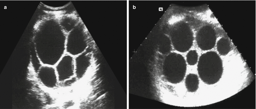

Fig. 26.5
Hepatic cystic hydatidosis (daughter cyst type). (a, b) Ultrasound demonstrates dark areas of small sphere-shaped daughter cysts with different sizes in the mother cyst, with their echo being lower than that of the mother cyst fluid
Calcification Type
Hydatidosis with a long illness course is demonstrated with thickened and rough external cyst wall and calcium deposition. By ultrasound, it is demonstrated as spotlike strong echoes at the hydatid cyst wall. In the cases with partial calcification, the sonogram shows arch-shaped light bands with strong echoes. In the cases with complete calcification of the hydatid cyst wall, the sonogram shows eggshell-like change, with obvious acoustic shadow at the lateral wall. Along with the severity of calcification, the acoustic shadow at posterior cyst is increasingly obvious, indicating decline or even death of the hydatid.
Consolidation Type
During the long illness course of hydatidosis, the cyst is gradually subject to degeneration, necrosis, lysis, and shrinkage into cheese appearance due to decreased and absorbed cystic fluid. In the cyst, consolidation mass is demonstrated with uneven intensity of echoes, which is typically gyrus like. The consolidated space-occupying lesion occurs due to collapse and folding of the mother cyst after absorption of cystic fluid as well as degeneration and necrosis of the daughter cysts. Its sonogram resembles to benign tumor with homogeneous echo.
Infective Type
Secondary infection occurs in the hydatid cyst to form pus, with collapse, necrosis, and decreased tension of the mother cyst. The mother cyst, therefore, deforms with unevenly thickened and coarse cystic wall. The internal cyst sheds off in the external cyst, which is twisted and irregular. The daughter cyst also deforms or is dried up, with uneven echoes of cotton-like light spots, light strips, and small light masses in different intensities filling up the cyst.
Rupture Type
The hydatid cyst sheds off from the external cyst wall, with collapsed, shrunk, and invaginated cyst wall that is twisted and folded, floating in the cystic fluid. In the liquid dark area, it is demonstrated with arch and folded cord-like strong echo band. When the posture is changed, the echo band may be floating and deformed.
X-Ray Radiology
Liver
The contour is enlarged. In the cases with giant hydatid in the right liver lobe compressing the right diaphragm, the diaphragm is observed to be elevated to cause limited breathing movement and even left shift of the heart. In children patients, the right lower thorax protrudes with widened intercostal space.
Calcification
Calcification can be found in the hepatic area. The ring-shaped, semiring-shaped, or eggshell-like calcification indicates cystic wall calcification. Intracystic calcification is demonstrated with round or round-like nodular calcification or stratified calcification.
Complications
Signs of hepatic abscess can be observed. The patients are demonstrated to have elevated right diaphragm, blurry diaphragmatic surface, and limited breathing movement. Otherwise, the complications may be demonstrated with right pleural inflammatory responses and effusion, blurry costophrenic angle and cardiodiaphragmatic angle, and poorly defined boundary between right lower chest and hepatic area. By pneumoperitoneography, left subphrenic inflation occurs due to right phrenic adhesion with no inflation.
Penetration Into Lungs
After infections occur to complicate hydatidosis at the top of right liver lobe, the hydatid may penetrate the diaphragm into the lung, which commonly causes pulmonary abscess at the anterior or posterior fundus segment in the right lower lung lobe. It is demonstrated as elevated right diaphragm or local protrusion of the right diaphragm, decreased breathing movement, inflammatory mass or lump in the right lower lung field, thickened pulmonary markings, and cord-like shadows extending toward the pulmonary hilum.
Penetration Into Thoracic Cavity
The hydatid cyst at the top of right liver lobe can penetrate into the right thoracic cavity to cause acute pleural effusion, with the lung compressed. In the cases with complicating bronchial fistula, hydropneumothorax can be demonstrated.
CT Scanning
By CT scanning, hydatidosis can be divided into cystic type, cystic calcification type, consolidation type, and mixed type. The former three types are basically corresponding to the germination, development, degeneration, and death of hepatic hydatid.
Cystic Type
In the liver, there are singular or multiple cysts that are round or round-like in shape with well-defined boundaries and smooth margins. Their densities are even and homogeneous, being close to the density of water. The cystic wall is thin and even, with no nodules. The intrahepatic bile ducts, vascular vessels, and adjacent organs are compressed with shifts. By contrast scanning, the cystic fluid and the cystic wall are demonstrated with no enhancement.
Simplex Cystic Type
In the liver, the cyst has smooth and sharp margin, which is singular or multiple round- or oval-shaped shadows with different sizes and even densities. The cystic wall is demonstrated as linear dense shadow in a thickness of 1–5 mm. By contrast scanning, no obvious enhancement is observed in the cyst (Fig. 26.6).
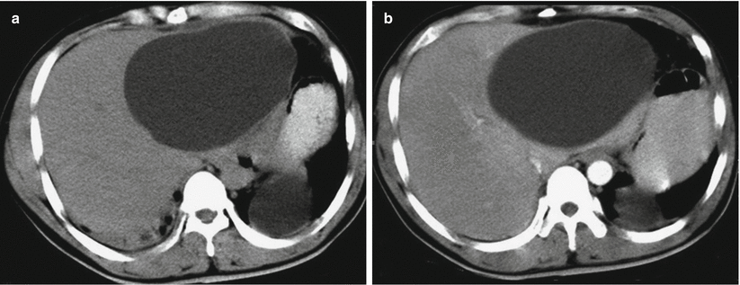

Fig. 26.6
Hepatic cystic hydatidosis (simplex cystic type). (a) Plain CT scanning demonstrates oval-shaped low-density shadow in the left liver lobe, with smooth and sharp margin and even density. The cystic wall is observed to be thin; (b) contrast CT scanning demonstrates no intracystic enhancement
Multiple Daughter Cyst Type
Emergence of multiple daughter cysts in the mother cyst is one of the characteristic manifestations of hepatic hydatidosis. There may be daughter cyst shadows in the mother cyst in different quantities and sizes and in round or irregular shape. The fibrous septum has different thicknesses. The daughter cysts are arranged around the mother cyst-like honeycomb or wheel, with their density being lower than their mother cyst. Along with increases of daughter cysts in quantity and size, the daughter cysts compress each other to be deformed into round shape, diamond shape, or polygon shape, which are distributed in the mother cyst-like grape cluster. The mother cyst fluid scatters between the daughter cysts, with higher density than daughter cyst fluid (Fig. 26.7). In the other cases, the daughter cysts are small in size and quantity. In the early stage, the small and round daughter cysts are arranged close to the mother cyst wall, with their density being close to the mother cyst (Fig. 26.8).
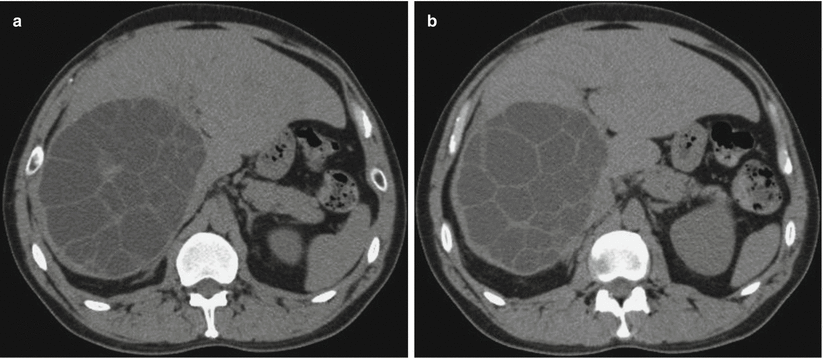
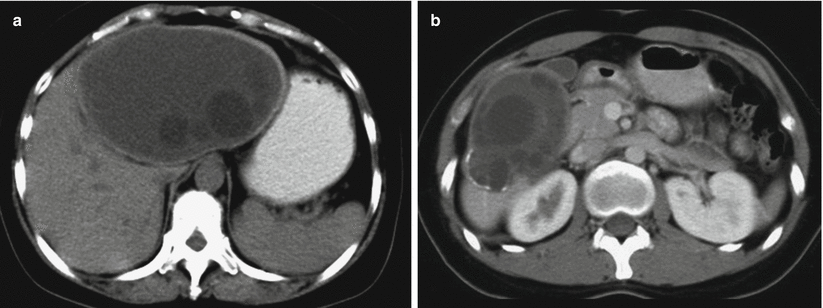

Fig. 26.7
Hepatic cystic hydatidosis (multiple daughter cyst type). (a, b) Plain CT scanning demonstrates hydatidosis at the right liver lobe, with multiple daughter cysts filling up the mother cyst that compress each other in honeycomb sign

Fig. 26.8
Hepatic cystic hydatidosis (multiple daughter cyst type). (a, b) Plain CT scanning demonstrates multiple round low-density shadows in the liver, with internal multiple lower-density round-like shadows of daughter cysts. The shadows are well defined, with smooth and sharp margins. Calcification can be observed at the posteroexterior margin of the lesion in the right liver lobe. By contrast scanning, no obvious enhancement is observed
Secondary Infection Type
The cystic wall is subject to irregular thickening, with poorly defined boundaries. In some cases, the cystic wall is imperfect, with increased but uneven intracystic density. The boundary of the cysts is poorly defined, sometimes with fluid level observed. By contrast scanning, the thickened cystic wall can be demonstrated with enhancement.
Rupture Type
David further divided the rupture type into two subtypes, internal cyst rupture and direct rupture. The subtype of internal cyst rupture is demonstrated with ruptured internal cyst but with its content within the external cyst. The cystic fluid gains its access into the external cyst and the internal cystic wall stripped partially or completely off the external cyst. By CT scanning, it is demonstrated as cyst bilateral sign or ribbon sign with wavy internal cyst membrane in the cystic lumen (Fig. 26.9). The completely separated internal cyst is subject to collapse and curling like a small lily. The subtype of direct rupture refers to rupture of both internal and external cysts, with the cystic fluid and their contents being directly drained into the abdominal cavity, ?? cavity or position inferior to the hepatic capsule. At this time, the hydatid cyst collapses and is deformed, with effusion at the abdominal cavity and the hepatic periphery as well as the daughter cyst shadows (Figs. 26.10 and 26.11). The rupture of hydatid cyst at the top of liver may cause intrathoracic complications or wrapping pleural effusion.
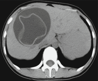
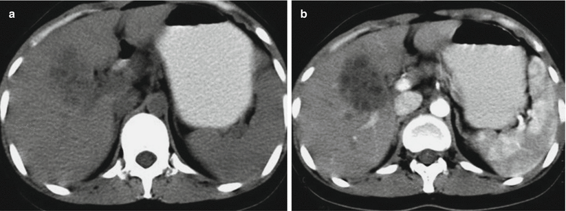
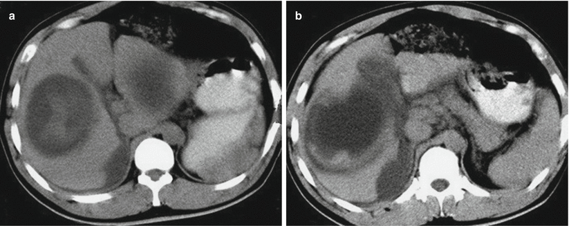

Fig. 26.9
Hepatic hydatid rupture. Plain CT scanning demonstrates collapse and shrinkage of the ruptured hydatid cystic wall, which is floating in the cystic fluid to form typical ribbon sign

Fig. 26.10
Hepatic hydatid rupture. (a, b) Plain CT scanning demonstrates increased uneven density of intrahepatic cyst with poorly defined boundary. The intrahepatic bile ducts around the lesion are demonstrated to be slightly dilated. The lesion is not enhanced by contrast scanning

Fig. 26.11
Hepatic hydatid rupture. (a, b) Plain CT scanning demonstrates cyst rupture in the right liver lobe. The lesion has poorly defined boundary. In the cystic lumen, the density is increased. In the gallbladder fossa and around the liver, effusion is observable
Cystic Calcification Type
Based on the position of cyst calcification, cystic calcification can be further divided into two subtypes, cystic wall calcification and intracystic calcification. Cystic wall calcification is arch-shaped continuous or continual calcification along the cystic wall. Otherwise, the calcification may be shell-like or spotlike calcifications. The cystic wall is demonstrated to be thickened, with even intracystic density that is close to water density. Sometimes, gas-fluid level can be observed in the cyst, which shows no enhancement by contrast scanning. Intracystic calcification is demonstrated with patches or spots of calcification shadows or intracystic linear high but even density shadow. The cystic fluid has high density, but with no enhancement by contrast scanning.
Consolidation Type
Due to the long illness course, the hydatid shows decreased activity to result in degeneration and necrosis. The absorption and condensation of cystic fluid cause caseous change, which is mixed with softened internal cyst or daughter cyst to increase the CT value. The CT value is also uneven, being close to that of consolidation tumor density shadow.
Mixed Type
In just one case, at least the above two types of lesions coexist, with CT scanning demonstrating characteristic lesions of different types.
Stay updated, free articles. Join our Telegram channel

Full access? Get Clinical Tree


