53 Ilioinguinal Nerve Block
The lower abdominal wall is primarily supplied by the subcostal, iliohypogastric, ilioinguinal, and genitofemoral nerves. The last three nerves arise from the lumbar plexus. Blocks of the ilioinguinal nerve are often performed to provide postoperative pain relief from inguinal hernia repair or for selective diagnostic purposes. Because transversus abdominis plane (TAP) blocks often fail to provide analgesia in the L1 distribution, ilioinguinal blocks may be of greater benefit for inguinal surgical procedures.1
The ilioinguinal nerve emerges between the muscles of the abdominal wall. The longest running course of the nerve is between the internal oblique and transversus muscles. The iliohypogastric nerve has a parallel course to the ilioinguinal nerve, running cephalad (superior) and medial. Of the three abdominal wall muscles (external oblique, internal oblique, and transversus), the internal oblique is usually the thickest.2
Suggested Technique
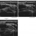
Nerve and muscle visibility are best cephalad to the pelvic brim.3 In the classic location for ilioinguinal block (2 cm medial and 2 cm superior to the anterior-superior iliac spine), the external oblique muscle is often aponeurotic, and therefore it is difficult to visualize this layer over the nerves. The first step is to obtain a view of the three abdominal wall muscles (external oblique, internal oblique, and transversus) to identify the ilioinguinal nerves between the internal oblique and transversus. Alternatively, the deep circumflex artery can be followed up from the external iliac artery until it meets the ilioinguinal nerves.
Key Points
| Ilioinguinal Nerve Block | The Essentials |
|---|---|
| Anatomy | The ILIH nerves lie between the IO and TA near the base of the IC. |
| The ILN lies 6 mm medial to the IC. | |
| The IHN lies 10 mm medial to the ILN. | |
| The IC has an inverted U appearance. | |
| The iliacus muscle lies under the TA at the IC. | |
| The DCIA is a recurrent branch of the external iliac artery. | |
| The ILN is often accompanied by the DCIA. | |
| The course of the ILN parallels the inside of the pelvic brim. |