V1, V2 and V3 – Trigeminal nerves – first, second and third divisions
MHz – Megahertz
USG – Ultrasonography
NSAIDs – Nonsteroidal antiinflammatory drugs
HIV – Human immunodeficiency virus
HBsAg – Hepatitis B virus antigen
HCV – Hepatitis C virus
NBM – Nil by mouth
IV – Intravenous
IHC – Immunohistochemistry
mAs – Milliampere second
Introduction
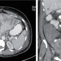
Head and neck region is complex in terms of the multitude of structures located in a fairly compact location and hence a complex variety of presenting pathologies. It is a pathway for numerous important structures traversing from the head to the rest of the body, which need to be differentiated from the normal structures. However, accurate cross-sectional imaging modalities such as CT or MRI may proclaim; obtaining a tissue sample is vital to provide a histopathological diagnosis (in tumours) or causative organisms (in infective pathologies) for further treatment.
Majority of the lesions in the region of head and neck are palpable. These may be amenable to FNAC or biopsy. However, the lesions may not be adequately sampled. And there are risks of inadvertent passage of the needle through important structures along the needle tract, especially vascular structures which may result in profuse bleeding. Hence, image guidance is a superior method to perform FNAC or biopsy accurately and safely not only in palpable lesions but also those which are deep-seated and hence not palpable.
The image guidance may be provided by the following:
• Fluoroscopy: This is rarely utilized except for direct approach to cervical vertebral osseous pathologies especially involving the posterior neural arch and transpedicular approach towards vertebral body lesions.
• Ultrasound (USG): This is preferred for providing guidance for superficially located lesions such as the thyroid and most cervical lymph nodes. It provides real-time guidance and is relatively safe but requires skillful hands and a steep learning curve.
• CT: This is excellent for providing guidance during sampling of the deep-seated lesions and those inaccessible to USG. The trajectory selected needs to be extremely precise owing to the variety of critical structures potentially encountered. The technique also usually requires general anaesthesia. If CT fluoroscopy is available, it also provides real-time guidance.
• MRI: Although MRI provides excellent soft-tissue contrast, it is extremely difficult to get access to structures in a closed bore high-field MRI magnet. Biopsies may be possible with open magnets which lack in resolution. MR-compatible anaesthesia equipment and other instruments are also a requirement during the entire process.
Hence, currently MRI guidance for guided biopsies is usually reserved for breast and prostate.
Patient selection
Certain questions need to be answered by every interventional radiologist before a patient is subjected to an image-guided procedure as it is a daunting procedure for most patients to undergo.
• Is there a real need to do the biopsy or can previous imaging or additional appropriate imaging be better?
• Although a lesion location makes it inaccessible to blind biopsy and too risky for other modes of biopsy, but is it amenable to appropriate image guidance for performing the biopsy?
• If multiple lesions are present, which one should be targeted? Which is most accessible biopsiable lesion under due risk?
• Is the biopsy going to change the course of patient management? If the diagnosis is going to be decisive for surgical intervention vs other forms of therapy or benign vs malignant, only then a biopsy needs to be performed.
• Are we dealing with a “no touch lesion” – vascular lesions (juvenile nasopharyngeal angiofibroma), fibrous dysplasia and asymmetric appearances of pterygoid plexus are such typical lesions which should not/need not be biopsied.
• Is the patient going to be able to lie still during the entire procedure? Are there any complications to be anticipated and prepared for before biopsy?
Preprocedure protocol
This is most important in the entire process of any image-guided intervention such as FNAC, biopsy or aspiration/drainage to select appropriate patients, plan the approach, avoid inaccurate sampling, prevent complications and provide the most representative tissue to make diagnosis possible.
1. Selection of the procedure and nature of guidance: Relevant procedure must be chosen with respect to FNAC vs biopsy as well as use of CT vs ultrasound for approaching the lesion to be sampled. These are detailed in the following.
2. Review of previous imaging: Review of all available prior imaging is essential. If a PET-CT is available, the area showing high uptake is usually the one to target if it is safe and feasible. If not available, sampling the solid enhancing portion of the lesion gives better yield. If recent study is not available, a new study may be insisted upon. If unclear from the prior imaging, intravenous contrast for CT-guided procedures may be administered to identify vascular structures in the lesion vicinity.
3. Consent and preprocedure counselling:
The patient must be informed details of the procedure in the language he/she understands so that an informed consent (legal) is provided and also entails good patient cooperation. In cases where the patient is unable to cooperate, then an anaesthesiologist must be on board to provide appropriate sedation or anaesthesia with further appropriate consenting. Any allergies must also be proactively sought and documented beforehand.
4. Review of medications being used by patient and laboratory investigations:
It is imperative to know if patients are on medications such as anticoagulants/aspirin. Depending on the drug used, these need to be temporarily stopped prior to the procedure. Low-molecular-weight heparin may be discontinued 24 h prior to the procedure and resumed 24 h postprocedure. Antiplatelet drugs such as clopidogrel need to be discontinued at least 5 days prior to the biopsy.
Usually, temporary cessation will be required for biopsies of deep-seated lesions and not for superficial FNACs. Concomitant risk factors must be also assessed, viz., history of coagulation disorder, platelet deficiency, liver dysfunction, immunocompromise, septicaemia, etc.
Laboratory investigations: Appropriate investigations may need to be requested and checked. These include HIV/HBsAg/HCV status, coagulation parameters (including bleeding time, clotting time, prothrombin time or INR as appropriate and platelet count) and tests as required by anaesthesiologists for safe administration of anaesthesia.
5. Anaesthesia: Sedation, local (superficial) vs general (deep seated) – the nature and location of the lesion, age and patient cooperation primarily influence whether anaesthesia is required and if so the appropriate choice of the suitable type. Sedation using fentanyl or midazolam is usually enough with general anaesthesia for uncooperative patients, children or lesions in critical or deep-seated locations.
Anaesthesia appointment: Most departments require proper advanced planning of appointments for procedures to be performed under sedation or anaesthesia as per protocols decided by the Department of Anaesthesia.
6. Nil by Mouth (NBM) status: This may be required for approximately 4–6 h if intravenous contrast or anaesthesia need to be administered.
7. Planning of actual procedure (tailored to each patient/lesion):
(a) Trajectory: Optimal trajectory needs to be planned depending on the site of lesion and details of the structures encountered along the planned trajectory.
(b) Patient positioning: The patient positioning (e.g. supine/prone/decubitus) has to be planned with respect to the expected trajectory. It is a prerequisite for the patient to be able to provide appropriate position with respect to biopsies especially of deep-seated lesions under CT guidance.
(c) Appropriate instrumentation (types of needles for biopsy and other instruments): This needs to be wisely chosen as they are not only expensive, but also the use of multiple changes in the devices used entails possibility of more complications.
(d) Optimum medium: The medium for sending the sample must be aptly selected after confirming with the pathologist/microbiologist: Formalin for tissues, normal saline for undetermined use to be selected later, culture tubes with or without media for collection of infective samples to be used for culture/growth. These must be planned beforehand to avoid wastage of obtained tissue samples and repeat procedures.
8. Preprocedure medications: An IV line must be secured especially if deep-seated or complex lesions are planned to be targeted for delivery of intravenous drugs. As there are no consistent guidelines on preprocedure medications in head and neck biopsies, the departmental policies can be decided locally. One must also enquire regarding any allergies to medications especially those which may be used in case of emergency/unexpected complication.
9. Actual procedure of image-guided FNAC/biopsy – This is explained in the following respective subsections.
10. Postprocedure confirmation: In case of FNAC, it is wise to confirm adequacy of aspirate by a cytopathologist to avoid repeat “passes”. It may not always be possible to assess adequacy on biopsy samples. But a discussion with the treating team when a certain diagnosis (such as lymphoma) is under consideration enables adequate sampling (for example, IHC, marker or receptor studies).
Postprocedure protocol
This is also an important part of any image-guided procedures to ensure the safety of the patients as well as safeguarding the interests of the performing radiologist. This protocol must be decided by individual department and diligently followed.
The patient is usually observed for about 2 h (lesser if a simple superficial lesion is biopsied and procedure was uncomplicated) before discharge or sending back to the ward or home. It is also a good practice to follow to inform the concerned referring consultant about the successful performance of the biopsy and any untoward complications that may have occurred and if admission of the patient is necessary for the same.
At the time of discharge, patient must receive explicit knowledge about postprocedural care including possible late complications, how to recognize them and appropriately guide preliminary care and further access to appropriate management. Analgesics and local application of anaesthetic spray may be required in some patients. Most patients do not require antibiotics. The patients also need to be advised when to resume medications if they have been discontinued prior to the guided procedure.
Further follow-up with clinical colleague and imaging if necessary may also need to be informed.
Ultrasound-guided FNAC/biopsy
The supreme advantage of ultrasound for guidance is “real-time” visibility of the lesion to be sampled and all the structures in the trajectory including the needle itself. Real-time colour Doppler is also available to judge the vessels in the vicinity (Table 3.41.1).
Advantages and Disadvantages of Ultrasound in Guided Interventions
Advantages
Disadvantages
• More real-time capability
• Operator dependence
• No IV contrast required
• Learning curve in coordination of both hands in performing the procedure
• Portability
• Inability to visualize posterior to the bone/air containing structures and metal
• No radiation exposure
• Less cost
Ultrasound is ideally suited for FNAC and biopsy of superficially locations such as thyroid, parotid and nodal lesions.
How to approach a patient for ultrasound-guided interventions?
1. Selection of patients: In ultrasound-guided procedures, the lesion must not be deep to bone, calcification, air or metallic implants. A suitable acoustic window is a prerequisite as also matching of depth of lesion, length of needle used and depth of penetration by the USG probe used. Ultrasound probe itself is useful to apply pressure to reduce depth of overlying tissues, displace obstructing structures and even partially immobilize mobile lesions (such as lipomas) to make FNAC/biopsy easier.
2. Review of previous investigations especially cross-sectional imaging for optimal ultrasound guidance: Prior imaging is primarily useful to know the nature (solid vs cystic) of the lesion, its vascularity and structures in the vicinity of the trajectory.
Preprocedure workup is common to all image-guided procedures as described before:
Only gold members can continue reading. Log In or Register to continue