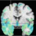
Fig. 1
Coronal Cine MRI slices visualizing the peristaltic wave in the descending colon
In order to integrate such a novel imaging technique into the daily clinical routine, it is mandatory to develop computer based analysis approaches. The huge amount of data which is acquired per patient needs automatic and semi-automatic methods in order to support the clinical expert in findings and diagnosis. Our main objective in this work is to model and quantify the activity of the large bowel hopefully providing the means to define the clinical significance of a variety of motility disorders in a wide range of patients. Necessary steps in the image processing workflow are defined, and technical approaches towards a computer aided diagnosis tool are proposed.
In the following, we present our preliminary results on a set of experimental data. Volunteer data was acquired and processed using the imaging techniques and algorithms presented in the next Sections.
2 Image Acquisition using Cine MRI
The volunteers undergo functional Cine MRI. The standardized functional Cine MRI examination is performed at 6 AM, after a minimum starving phase of 8 hours, on 1.5-T system (Siemens Avanto). The volunteers are examined in supine position. Neither premedication nor contrast agent is applied. The dynamic part of the examination consists of 2 blocks of repeated measurements covering the entire abdomen, using a T2-weighted HASTE-sequence. Each block contains a stack of approximately 200 slices over time orientated in coronal plane adapted to the anatomic course of the descending colon (see Fig. 1). The image resolution is 256 × 320. Between the 2 dynamic blocks of measurements a prokinetic agent is administered in order to stimulate the colon motility. For the further analysis of the peristaltic motion, the subsequence (usually about 20 slices) showing this motion is manually selected from the image blocks. The pre-scan without stimulation was up to now only used in our previous studies [6, 7].
In order to achieve the fastest possible frame rate for our MRI acquisition, no respiratory gating can be used during the scans. The time between two successive frames could be reduced to approximately 1.4 seconds which is fast enough to visualize the peristaltic wave. Still, the sequences suffer from breathing motion artifacts which makes the identification of corresponding points in the colon quite hard. This was not a problem in our previous studies where manually extracted diameters were measured over time and stimulated and non-stimulated sequences were compared, the identification of corresponding points could be achieved manually by the medical expert. In order to automatize such procedures, there is need for an accurate motion compensation in a post-processing step. This leads us to the first step in the image processing chain (see Fig 2).


Fig. 2
Our image processing chain within a computer aided diagnosis tool for colon motility dysfunctions
3 Image Processing
In order to develop a computer aided diagnosis tool for colon motility dysfunctions, we first identify necessary steps within the image processing chain. We already mentioned the problem of breathing motion artifacts in the Cine MRI data sets. Once, these artifacts can be successfully removed, all further steps will benefit from the motion compensation. Our later analysis of the motility is based on the segmentation of the colon in all slices over time. We propose a semi-automatic approach based on interactive graph-cuts. This will be explained in more detail in Sect. 3.2. The actual analysis and our approach for extracting clinically valuable measurements is then described in Sect. 4. The full image processing chain is sketched in Fig. 2.
3.1 Motion Compensation
As already mentioned, no respiratory gating techniques are used during the image acquisition in order to achieve a high frame rate. The resulting breathing artifacts in the image sequences are visible in a vertical jumping of the abdominal organs effected by breathing motion such as liver, kidney, and of course the colon itself. For our further processing steps, a compensation of this motion is of great interest. In order to make the identification of corresponding points in the colon much easier we try to stabilize the image parts of interest. We propose a semi-automatic motion compensation method. Therefore, the user selects a subregion in one of the slices, which we call the reference frame. The selected region should represent the overall breathing motion but should not include any parts of the colon. So we can avoid to eventually compensate for colon motility. Within the selected region features are extracted which fulfill the Harris [10, 14] assumptions. The robust and fast implementation of the Lucas-Kanade optical flow method [3] is then used to compute the displacements of each single feature in every frame in respect to the reference frame. Afterwards we compute a mean vertical displacement for every frame which represents the overall breathing motion. By translating each frame by its corresponding mean displacement we can compensate for this motion. In practice, the image part containing the lower liver boundary and right kidney turned out to be a good region for tracking the breathing motion (see also Fig. 3). These parts show a very similar breathing motion such as the colon itself. The result of the motion compensation is a clear stabilization of all organs with a similar movement. Naturally, former stable parts (e.g. the spine, see also Fig. 3d) are consecutively jumping within the image series, However, this fact does not present a problem for the further processing of the colon.
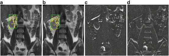

Fig. 3
(a) Selected region and extracted Harris feature points. (b) Tracked features in one of the following frames. (c) Difference image between reference frame and consecutive frame before compensation, (d) and the difference image after compensation. Clearly visible, the motion at the lower boundary of the liver has been reduced
3.2 Colon Segmentation
The segmentation of the colon is crucial for our further analysis. Shape and appearance have to be well preserved by the segmentation. The individual patient’s anatomy is particularly reflected in varying orientation and form of the large bowel. A segmentation method should be highly flexible to handle these variations. To this end, we use the interactive graph-cuts approach proposed by Boykov and Jolly [4]. The segmentation is defined as an energy formulation

where A indicates a segmentation of the pixels x of domain Ω of the image series I into two subsets  (object pixels) and
  (background pixels) with

(1)
 (object pixels) and
  (background pixels) with

